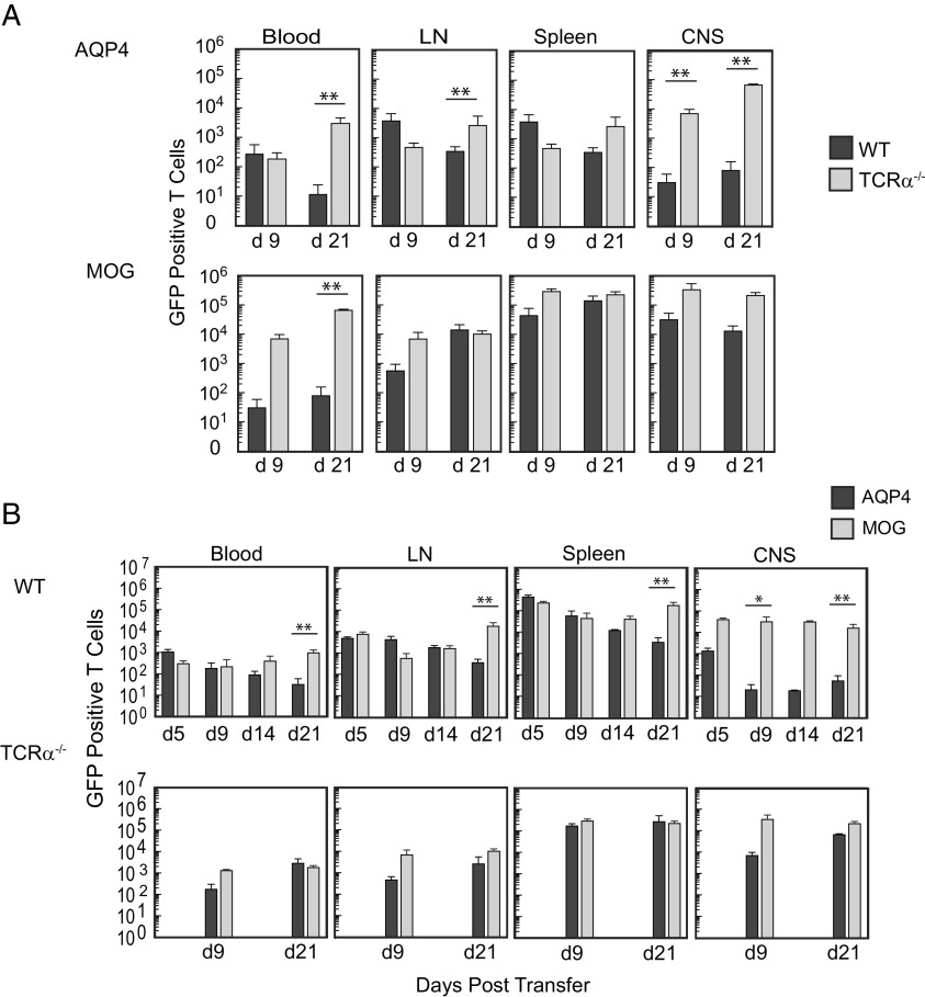
Fig. 6.
Pathogenic AQP4-specific Th17 cells persist in T cell–deficient mice but not in WT mice. (A) Donor Th17-polarized AQP4 p133–149-specific and MOG p35–55-specific primary T cells from actin-GFP/AQP4−/− mice were transferred into WT or T cell–deficient (TCRα−/−) mice and then examined for recovery in 100 μL blood, axillary and inguinal lymph nodes combined (LN), the spleen, and the CNS at 9 and 21 d by flow cytometry. Cells were gated on single, viable GFP+CD4 T cells. Counting beads were used for quantification. (B) AQP4 p133–149 or MOG p35–55-primed Th17-polarized LN cells from AQP4−/− actin-GFP+ mice were transferred into WT or TCRα−/− mice and then examined by flow cytometry at the indicated timepoints. Data shown for A and B represent a composite from three experiments [n = 6 per condition (mean ± SEM)]. Statistical comparison of surviving numbers of GFP+ donor cells was performed by the Mann–Whitney U test (**P < 0.01).

