Fig. 1.
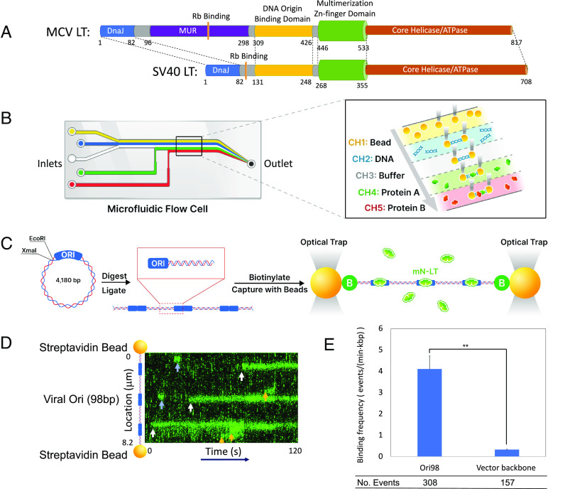
MCV LT specifically binds to the MCV replication origin. (A) MCV and SV40 LT helicase oncoprotein domains. MCV and SV40 LT are homologous helicases sharing DnaJ, retinoblastoma (Rb) protein-binding, DNA origin binding (OBD), multimerization Zn-finger and helicase domains. MCV LT contains an MCV unique region (MUR) that is not present in SV40 LT. (B) Microfluidic chip for DNA capture. (B, Left) Schematic flow cell. (B, Right) Details of the laminar flow channels. Initial DNA-polystyrene bead capture occurs in channel 1, followed by DNA tethering in channel 2, DNA quality check in PBS buffer in channel 3, and protein binding and imaging in channels 4 and 5. (C) Cloning of biotinylated Ori98 DNA. pMC.Ori98 Plasmids were digested at XmaI/EcoRI sites and self-ligated to form random origin multimers (1× to 7×), and CG ends were filled with biotinylated dCTP and dGTP using Klenow fragment. The number and location of Ori98 sequences were determined from DNA length in each assay. (D) Representative kymograph of mN-LT bound to multimeric pMC.Ori98 (3×) DNA showing both prolonged and transient binding events. Three origin (white arrows) and three non-origin-binding events (orange arrows) are shown. Transient binding events (< 5 s duration, blue arrows) were not included in subsequent analyses. (E) Ori98 sequence has high binding specificity compared to non-Ori98 pMC.BESPX backbone DNA sequence. Binding frequency for Ori98 and non-Ori98-binding events were collected from 30 DNAs, 5 min exposure each. Statistical analysis was performed using an unpaired t test, P = 0.0085.

