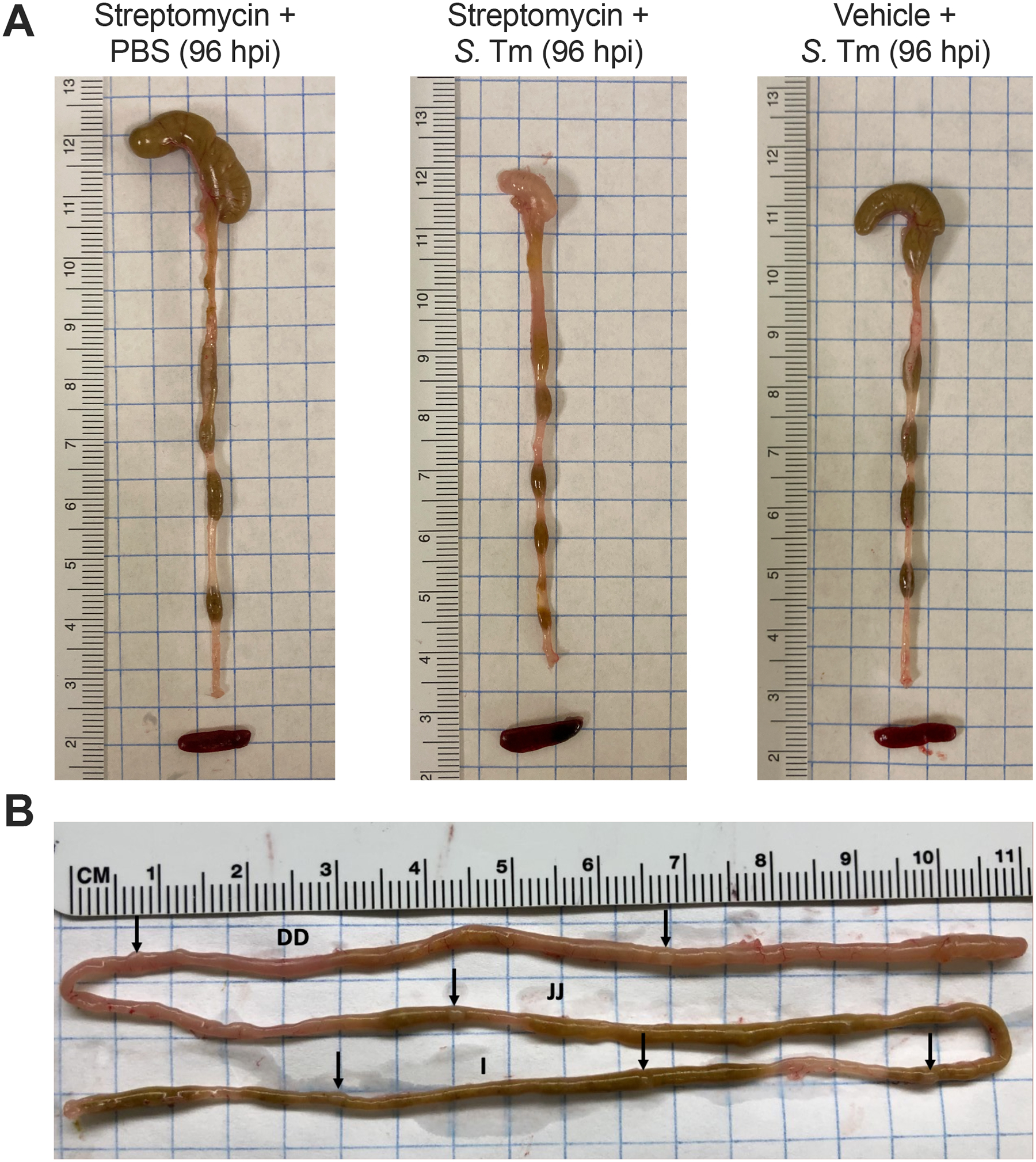
Figure 1. Representative gross pathology in the streptomycin-colitis model of S. Typhimurium infection.

(A) Gross pathology of the cecum, colon, and spleen collected from C57BL6/J mice pretreated with 20 mg of oral streptomycin and mock-infected with PBS, or inoculated intragastrically with 1×10⁹ CFU of S. Typhimurium IR715 (S. Tm) for 4 days following pretreatment gavage with streptomycin or vehicle (sterile water). The small, thick cecum devoid of content is a hallmark of the inflammatory response triggered by S. Typhimurium in mice pretreated with oral streptomycin. (B) Representative photograph of small intestine on day 4 of Salmonella infection. Black arrows indicate the Peyer’s patches, which are easily identified by their protruding appearance on the anti-mesenteric site. I, ileum; JJ, jejunum; DD, duodenum.
