
Figure 2. Representative H&E-stained ceca from uninfected mice or 96 h after intestinal S. Typhimurium infection.
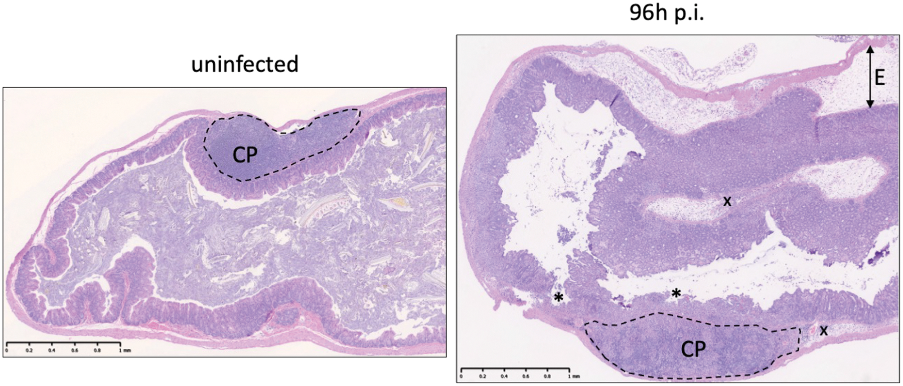
The dashed line indicates the cecal patch (CP), which shouldn’t be confused with tissue inflammation. Asterisks (*) indicate severe ulceration of the epithelial lining. The arrow indicates severe submucosal edema (E). The x indicates submucosal infiltration of immune cells. 60 x magnification, scale bar 1mm.