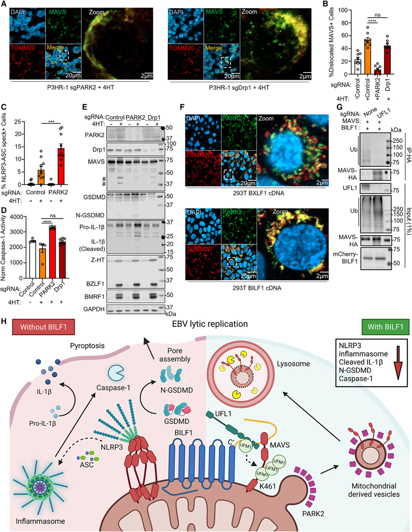
Figure 7. BILF1 dislocates MAVS to mitochondria derived vesicles.
(A) Immunofluorescence analysis of MAVS and TOMM20 in P3HR-1 expressing the indicated PARK2 or Drp1 sgRNAs and 4HT-induced for 24 h.
(B) Mean ± SEM percentage of cells with dislocated MAVS from n = 3 replicates, as judged by appearance of MAVS puncta that did not overlap with TOMM20 signal as in (A), using data from 25 randomly selected panels of 500 nuclei, analyzed using ImageJ ComDet plugin.
(C) Mean ± SEM percentage of P3HR-1 with NLRP3/ASC specks from n = 3 replicates, using data from 20 randomly selected panels of 200 nuclei, analyzed by ImageJ ComDet plugin.
(D) Mean ± SEM caspase-1 activity normalized by live cell number from n = 3 replicates of P3HR-1 expressing the indicated sgRNA and 4HT-induced for 24 h, as indicated.
(E) Immunoblot of WCL from P3HR-1 expressing the indicated sgRNA and 4HT-induced for 24 h. # denotes low molecular weight bands immunoreactive with anti-MAVS antibody. Representative of n = 3.
(F) PAKR2 and TOMM20 immunofluorescence analysis in 293T transfected with BILF1 or BXLF1 cDNA for 24 h.
(G) Immunoblot of 1% input vs. anti-HA-MAVS immunopurified from wild type or UFL1-KO 293T transfected with MAVS and BILF1 cDNA for 24 h, as indicated. Representative of n = 2.
(H) Schematic of NLRP3 inflammasome subversion by BILF1. BILF1 recruits UFL1 to mediate MAVS UFMylation, which together with PARK2 triggers selective MAVS removal from the mitochondrial outer membrane, MDV packaging and delivery to lysosomes, preventing NLRP3 inflammasome activation and pyroptosis. Student’s t test was performed, with ****p < 0.0001. ***p < 0.001. ns > 0.05. See also Figure S6.

