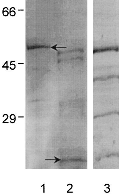
FIG. 2.
SDS-polyacrylamide gel electrophoresis of expressed 50-kDa recombinant E. canis P28-thioredoxin fusion protein (lane 1, arrow) and 16-kDa thioredoxin control (lane 2, arrow) and corresponding immunoblot of recombinant E. canis P28-thioredoxin fusion protein recognized by convalescent-phase E. canis canine antiserum (lane 3). The thioredoxin control antigen did not react with the E. canis antiserum (not shown). Numbers on the left are molecular masses in kilodaltons.

