Figure 2.

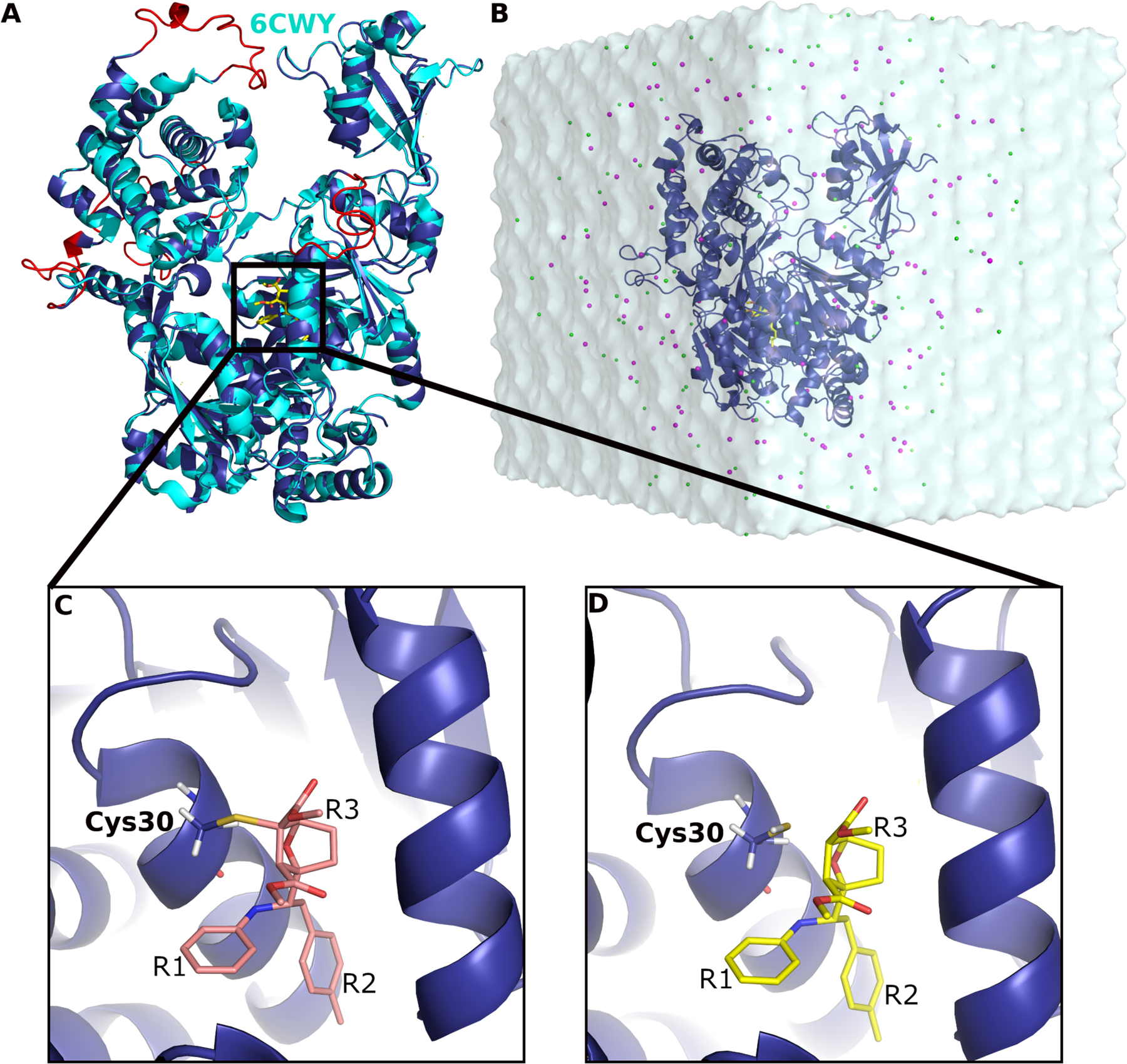
(A) Computational model of SUMO E1 (blue cartoon) aligned to the X-ray structure (cyan cartoon) in complex with COH000 (yellow sticks). The missing regions added through homology modeling are highlighted in red. (B) LiGaMD simulation input system with COH000 (yellow sticks) bound to SUMO-E1 (blue cartoon) that was solvated in 0.15 M NaCl (magenta and green beads) solution. (C) A covalent bond was formed between COH000 and Cys30 in SUMO E1 in the X-ray structure. (D) The protein-ligand covalent bond was broken for LiGaMD simulations of non-covalent COH000 interacting with the SUMO E1. The R1, R2 and R3 groups are indicated in (C) and (D).
