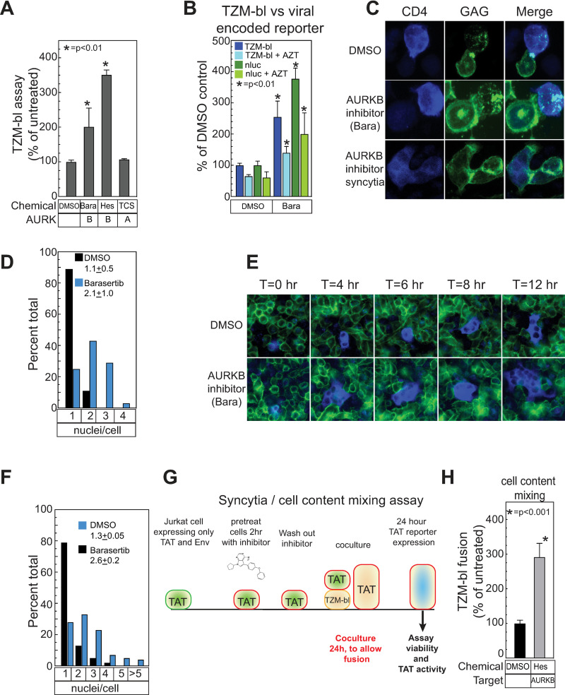
Fig 5. AURKB specifically regulates the fusion activity of the HIV envelope through the cytoplasmic tail domain of Env.
(A) The effects of kinase inhibitors targeting AURKB (Barasertib, 20 μM, and Hesperidin, 5 μM) and AURKA (TC-S 7010, 10 μM) was determined as described in Fig 4. (B) HIV-1 Jurkat producer cells treated with DMSO only, 20 μM barasertib and/or 500 nM AZT as indicated were mixed with TZM-bl cells as described in Fig 4 and then assayed for the TZM-bl-encoded firefly luciferase or the virally encoded GFP-NanoLuc (Promega) reporter gene. Barasertib treatment was conducted as described in Fig 4 and the associated figure legend. AZT was added with barasertib and maintained at 500 nM throughout the experiment. (C) HIV producer cells (Jurkat) with a fluorescent protein (CFP) fused to HIV-gag and WT HIV-1 Env were treated for 2h with DMSO or barasertib (20 μM) and mixed with target cells (SupT1) expressing fluorescent CD4 (CD4-YFP) for 20 minutes, fixed, mounted and imaged by confocal microscopy with a 60X objective. Inhibition of AURKB resulted in elongated cell-cell contacts and the frequent formation of syncytia. (D) The distribution of nuclei per cell after treatment with the indicated inhibitors and cell mixing was quantified and normalized as a percentage by counting a minimum of 120 cells from 3 fields from independent experiments are plotted. Mean and SEM are indicated. (E) Selected time frames from live cell images of AURKB inhibitor (Barasertib, 20 μM, 2 hr) -treated Jurkat cells transfected with HIV with a fluorescent protein (CFP) fused to HIV-gag and WT HIV-1 Env seeded on a monolayer of HeLa cells expressing CD4-YFP. (F) The distribution of nuclei per cell 12 hr after treatment and mixing was quantified and normalized as a percentage by counting a minimum of 120 cells from 3 fields from independent experiments. Mean and SEM are indicated in red. (G) Schematic of TZM-bl based syncytia/content mixing assay. Jurkat cells were co-transfected with a plasmid expressing the HIV-1 envelope protein and a second plasmid encoding the HIV transactivator TAT. Since no genome or other viral replication proteins are present, no virions can be formed. The cells were resuspended in media containing kinase inhibitor and incubated for 2hr. Treated cells were then mixed with TZM-bl reporter cells (HeLa cells that express the HIV CD4 receptor and co-receptors and have a TAT responsive promoter upstream of the firefly luciferase reporter). At 24 hours, cell viability and the activity of the TAT-responsive promoter were assayed. Since no virions were produced, the only activation of the TAT-responsive promoter can occur with cell-to-cell fusion and contact mixing. (H) Jurkat cells expressing TAT and WT NL43 Env were treated with an AURKB inhibitor (Hesperidin, 5 μM) as described in (G) and membrane fusion with TZM-bl cells was measured by TAT reporter gene activity. The data shown are the average mean values obtained in an experiment performed with quadruplicate samples and are representative of three independent experiments. Error bars indicate the standard deviation of the data in all panels. P-values were calculated using a standard Student’s t-test and significant changes relative to DMSO treated controls are indicated.

