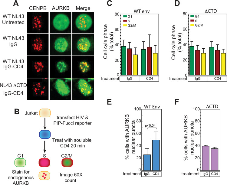
Fig 9. Interaction with soluble CD4 HIV Env is sufficient to relocalize AURKB.
(A) Jurkat cells were co-transfected with plasmids encoding the indicated HIV envelope, GFP-AURKB and mCherry-CENBP. 24 hours post transfection, cells were incubated with purified IgG or soluble IgG-CD4 fusion protein for 20 minutes, fixed, mounted and imaged at 60X by confocal microscopy. Treatment with the soluble CD4 fusion protein caused HIV to relocalized to CENBP adjacent foci. (B) Schematic for quantitation of CD4 induced AURKB localization and cell cycle changes. Jurkat cells were co-transfected with plasmids encoding the indicated HIV envelope, along with mAzurite-Histone H2B to mark the transfected cells and a plasmid that encodes the 2-color PIP-Fucci system than uses a YFP-PIP protein to mark G1 cells, an mCherry-Geminin to mark S-phase cells. G2/M phase cells are dual positive. 24 hours post transfection, cells were incubated with purified IgG or soluble IgG-CD4 fusion protein for 20 minutes, fixed, stained for endogenous AURKB with a far-red secondary (alexafluor 647), mounted and imaged at 60X by confocal microscopy. 10 independent fields were counted, and transfected cells were scored for CD4 induced changes to the cell cycle stage with (C) WT or (D) ΔCTD envelope. (E) Total AURKB localization at nuclear puncta by WT or (F) ΔCTD envelope. The data shown are the average mean values obtained from three independent experiments with a minimum of 550 cells counted per condition between experiments. Error bars indicate the standard deviation of the data in all panels. P-values were calculated using a standard Student’s t-test and significant changes of IgG treated control cells to soluble CD4 treated cells are indicated.

