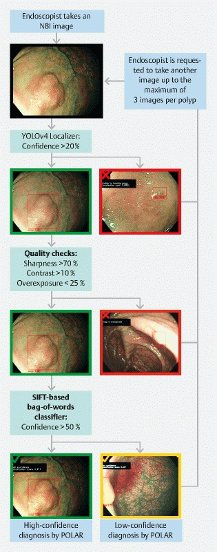
Fig. 1.

POLAR system design and user protocol during clinical validation. When using the POLAR system during clinical validation, the endoscopist has to take a maximum of three nonmagnified images using narrow-band imaging. After one image of the lesion is taken, it is processed by the POLAR system. If the system is able to provide a high-confidence diagnosis (a green mark is shown on the monitor), the endoscopist can continue with the procedure (i. e. resect the lesion). If the system is not able to provide a high-confidence diagnosis, the system provides feedback to the endoscopist on why this was not possible (e. g. not able to localize the lesion [red mark], not of sufficient quality [red mark], or only able to perform an optical diagnosis with low confidence [orange mark]). If the system is not able to provide a high-confidence diagnosis, the endoscopist has to take another image, up to a maximum of three per lesion. If the system is still not able to provide a high-confidence diagnosis after three images, the endoscopists can stop taking images, and proceed with the procedure. The low-confidence diagnosis with the highest prediction score is used as the final diagnosis of the system. If the system is not able to provide a low-confidence diagnosis after three images, this is considered as a failure of the system. NBI, narrow-band imaging.
