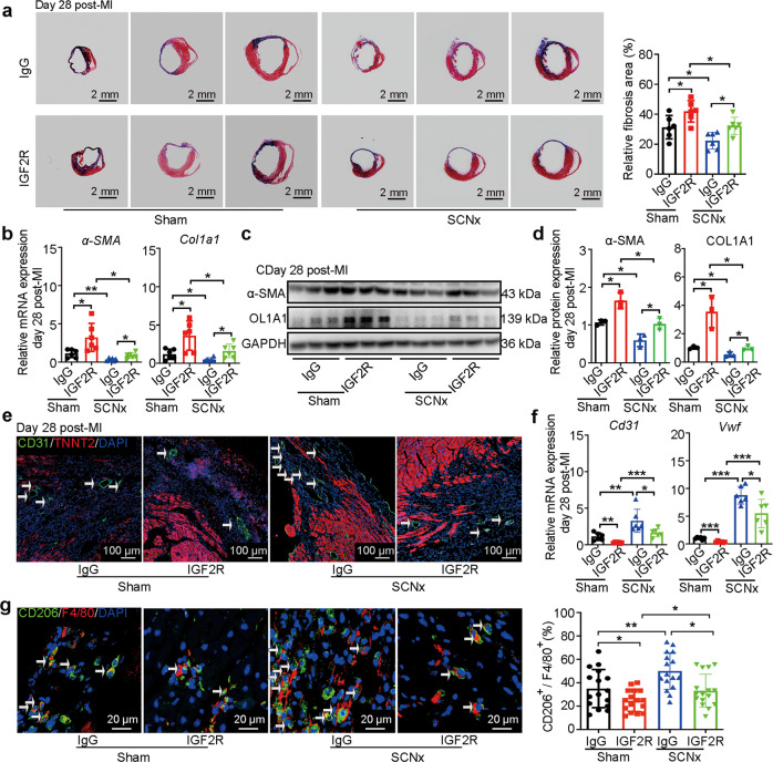
Fig. 7. IGF2R antibody treatment disturbed anti-fibrotic and anti-inflammatory effect of SCN ablation.
a Representative images of Masson’s trichrome-stained MI hearts, and quantification of fibrosis (% area) showing a significant increase in the fibrosis areas in IGF2R antibody-injected MI hearts (Scale bars: 2 mm, n = 6 per group). b The qRT-PCR analysis of α-SMAand Col1a1 expression as indicators of myocardial fibrosis (n = 6 per group). Western blot analysis (c) and relative densitometric quantification of α-SMA and COL1A1 (d) protein levels in MI hearts (n = 3 per group). e Representative images of immunofluorescence staining for CD31 and TNNT2, and quantification of angiogenesis in the border zone of the heart after MI (Scale bar: 100 μm). f The qRT-PCR analysis of Cd31 and Vwf in the infarction zone of MI hearts as indicators of the heart’s angiogenic ability (n = 6 per group). g Representative immunofluorescence staining images and quantitative analysis showing CD206+ and F4/80+ macrophages in the hearts of Sham-operated and SCNx mice on day 3 post-MI (Scale bars: 20 μm, n = 16 per group). Data are presented as mean ± SEM; two-way repeated-measures ANOVA; *P < 0.05, **P < 0.01, ***P < 0.001.

