



Abstract
Advances in instrumentation and technique have facilitated minimally invasive surgeries for cardiac myxoma treatment. This study aims to compare the clinical outcomes between the thoracoscopic and robotic approaches for myxoma resection. Intraoperative data and postoperative data of 46 patients who underwent either thoracoscopic (n = 15) or robotic (n = 31) cardiac myxoma resection in our center between July 2013 and September 2022 were retrospectively compared. There was no in-hospital death in either group. Meanwhile, the operative time and cardiopulmonary bypass time were significantly shorter in the robotic group than in thoracoscopic group (P = 0.015 and P = 0.035, respectively). Furthermore, shorter ICU stays (P = 0.006), shorter postoperative mechanical ventilation time (P = 0.035) and less thoracic drainage (P = 0.040) were observed in the robotic group. However, the operating room costs and total hospital costs were both significantly lower in thoracoscopic group (P = 0.004 and P = 0.007, respectively). There was no significant difference between two groups regarding the incidence of postoperative complications (P > 0.05). Lastly, a faster return to exercise was noted in robotic group than in thoracoscopic group (Log-Rank χ2 = 4.094, P = 0.043). Both approaches can be safe and feasible for myxoma resection. However, regardless of higher expenses, the robotic myxoma resection approach provides shorter operation time, less postoperative thoracic drainage, and faster recovery than total thoracoscopic technique.
Keywords: Myxoma, Minimally invasive surgical procedures, Thoracoscopy, Robotics
Introduction
Cardiac myxoma is the most common type of primary cardiac tumor [1], which accounts for about 75–80% of all the primary neoplasms in the heart [2]. The prevalence of the cardiac myxoma is estimated to be 0.5 per million per year [3]. Once diagnosed, surgical resection is usually recommended to prevent fatal complications such as valvular obstruction and embolization [4]. Since the first successful removal of left atrial myxoma in 1954 [5], sternotomy has been widely accepted as the standard procedure for cardiac myxoma resection [3]. However, minimally invasive techniques such as total thoracoscopic and robotic surgical approaches have evolved considerably during the last decade and obtained excellent outcomes [6, 7]. Thus far, the application of these minimally invasive techniques has expanded to mitral valve surgeries, congenital heart disease repair, atrial fibrillation ablation, coronary artery bypass grafting (CABG), and cardiac myxoma resection [8–11]. Nevertheless, compared with other diseases, reports on myxoma resection using either the robotic or total thoracoscopic technique is still relatively scarce, mainly due to the low incidence of myxoma and the limited number of hospitals equipped with the robotic system or thoracoscopic device [4].
Both the thoracoscopic and robotic approaches for myxoma resection have been associated with better visualization, smaller incision, and faster recovery than conventional sternotomy [12, 13]. Wei et al. previously compared the clinical outcomes between thoracoscopic and robotic surgeries for mitral valve repair [8]. They concluded that the thoracoscopic technique has more advantages in postoperative recovery and hospitalization expenses. However, research comparing the thoracoscopic and robotic procedures for myxoma resection has not been reported in any previous literature. Therefore, in this study, we sought to compare the clinical outcomes between these two approaches for myxoma resection. To the best of our knowledge, this study is the first of its kind reported in the literature.
Methods
Patient selection
A total of 46 patients who underwent minimally invasive cardiac myxoma resection in our center from July 2013 to September 2022 were included. Among these, 31 patients were treated using the Da Vinci Surgical System (robotic group), including 29 patients with left atrial myxoma (LAM) and 2 patients with right atrial myxoma (RAM). The other 15 cases were treated using a thoracoscopic procedure (thoracoscopic group), including 14 patients with LAM and 1 patient with RAM. Preoperative transthoracic echocardiography (TTE) was performed for each patient to evaluate the size, location, attached sites, and obstruction status of the tumors. The selection criteria for both the total thoracoscopic and robotic procedures were as follows: (1) without the requirement for concurrent procedures such as coronary surgery, aortic procedures, or aortic valve surgery; (2) without history of right thoracotomy, severe pericarditis, or severe pleuritis; (3)without thorax deformity or morbid obesity; (4) without peripheral vascular problems which may restrict femoral cannulation; (5) without history of asthma or chronic obstructive pulmonary disease (COPD). Furthermore, postoperative pathological examination of all subjects confirmed that all excised tumors were cardiac myxomas.
Robotic surgery technique
After double-lumen endotracheal intubation under general anesthesia, a transesophageal echocardiography (TEE) evaluation was performed. The patients were then maintained in supine position with the right thorax elevated to 30°. The cardiopulmonary bypass (CPB) was achieved via right internal jugular venous cannulation, right femoral arterial and venous cannulation. After the initiation of single-lung ventilation, a 3-cm working port was made on the anterior axillary line in the fourth intercostal space (ICS). The camera was inserted anteriorly at the same ICS. Two 8-mm endoscopic trocars were then placed at the anterior axillary line in the 3rd and 6th ICS for robotic arms. Meanwhile, the atrial retractor was inserted through the 5th ICS at the mid-clavicular line. After docking the Da Vinci robot (Intuitive Surgical, Sunnyvale, CA, USA), the pericardium was incised. Superior and inferior vena cava were sequentially blocked following the initiation of CPB. Cases with right atrial myxoma were directly excised via right atriotomy on the beating heart. For cases with left atrial myxoma resection, aortic blocking was completed using a Chitwood cross-clamp inserted through the 3rd or 4th ICS at the right mid-axillary line, while antegrade perfusion of cold-blood cardioplegic solution was administered. All the myxomas were excised completely. The defect of the atrial septum or atrial wall after myxoma resection was repaired by continuous suturing or using a pericardial patch when needed. TEE was required at the end of the procedure to assess if there was a residual tumor or interatrial shunting after septal reconstruction.
Thoracoscopic surgery technique
Three ports were made on the right chest wall. Port 1 (3–4 cm) was made at the 4th ICS of the right anterior axillary line, where the thoracoscope was inserted. Port 2 (2–3 cm) was made parasternally in the 2nd or 3rd ICS for entries of left-hand instruments. Port 3 (2–3 cm) was made in the 5th or 6th ICS lateral to the mid-clavicular line for entries of right-hand instruments. The establishment of the peripheral CPB and intra-thoracic part of the operation were the same as those of the robotic group. The operative scene of thoracoscopic and robotic myxoma resection is presented in Fig. 1.
Fig. 1.

Operative scene of robotic and thoracoscopic surgery. A Resection of left atrial myxoma (with arrow) during robotic surgery. B Resection of right atrial myxoma (with arrow) during thoracoscopic surgery
Data collection and follow-up
The operative parameters, in-hospital outcomes, and follow-up results between the robotic and thoracoscopic groups were compared. Peak pain score (Visual Analogue Scale, VAS) after surgery was observed to assess the postoperative pain. Clinical and follow-up data were obtained from electronic medical records and follow-up surveys. The patients were followed up at the outpatient center after discharge. A telephone interview was required for those who were lost to the outpatient center. In the follow-up, the time to return to exercise and the occurrence of any major adverse cardiovascular and cerebrovascular events (MACCE) were investigated for both groups. MACCE was defined as major adverse cardiovascular and cerebrovascular events since the day of operation, including events of cardiac death, acute coronary syndrome, new-onset arrhythmia, stroke, and peripheral vascular embolism.
Statistical analysis
Statistical analysis was performed using SPSS 25.0 (IBM, Chicago, Illinois, USA). Continuous variables with normal distribution were expressed as mean ± standard deviation (SD), and were compared by the Student’s t-test. Continuous variables with skewed distribution were expressed as median and 25–75 percentile, and were compared using the Mann–Whitney U test. Categorical variables were described as numbers and percentages, and were compared by Pearson’s Chi-square test, continuity adjusted Chi-square test or Fisher’s exact test. Survival data were analyzed using the Kaplan–Meier method and compared by log-rank test. A p-value of < 0.05 was considered statistically significant.
Results
There was no significant difference between two groups in terms of sex, age, BMI, cardiac function, blood creatinine, and parameters of preoperative echocardiography (Table 1). The incidence rates of various comorbidities, including coronary artery disease (CAD), diabetes, hypertension, embolism, and preoperative atrial fibrillation, were also similar between the two groups (Table 1).
Table 1.
Baseline characteristics
| Thoracoscopic | Robotic | P-value | |
|---|---|---|---|
| (N = 15) | (N = 31) | ||
| Gender (male/female) | 5/10 | 13/18 | 0.575 |
| Age (years) | 54.13 ± 11.26 | 53.10 ± 15.16 | 0.815 |
| BMI (kg/m2) | 22.74 ± 2.82 | 21.55 ± 3.10 | 0.219 |
| LVEF (%) | 63.73 ± 4.76 | 63.42 ± 3.34 | 0.797 |
| LAD (mm) | 40.00 ± 6.32 | 39.36 ± 6.12 | 0.744 |
| LVEDD (mm) | 47.71 ± 5.11 | 47.62 ± 3.69 | 0.944 |
| PASP (mmHg) | 35.0 (27.0,51.0) | 32.0 (28.0,46.0) | 0.851 |
| Blood creatinine (µmol/L) | 61.15 ± 16.12 | 59.10 ± 13.24 | 0.650 |
| Cardiac function [n (%)] | 0.933 | ||
| NHYA I/II | 9 (60.0) | 19 (61.3) | |
| NHYA III/IV | 6 (40.0) | 12 (38.7) | |
| Hypertension [n (%)] | 2 (13.3) | 3 (9.7) | 0.709 |
| Diabetes [n (%)] | 0 (0.0) | 2 (6.5) | 1.000 |
| Coronary artery disease [n (%)] | 0 (0.0) | 1 (3.2) | 1.000 |
| Embolism | |||
| Cerebral embolism [n (%)] | 2(13.3) | 6 (19.4) | 0.928 |
| Peripheral embolism [n (%)] | 0 (0.0) | 0 (0.0) | NA |
| Atrial fibrillation [n (%)] | 0 (0.0) | 0 (0.0) | NA |
BMI body mass index, LVEF left ventricular ejection fraction, LAD left atrial diameter, LVDD left ventricular end-diastolic diameter, PASP pulmonary artery systolic pressure, NYHA New York Heart Association functional class
The surgical data are presented in Table 2. All 46 operations were completed without transition to median sternotomy in either group. However, shorter CPB time (97.90 ± 24.48 vs. 118.27 ± 38.82 min, P = 0.035) and shorter operation time (225.39 ± 33.83 vs. 297.13 ± 98.70, P = 0.015) were observed in robotic group. Left atrial myxomas were more often excised via left atriotomy or biatriotomy when performing the robotic procedure. In contrast, left atrial myxomas were more frequently resected via right atriotomy when performing the thoracoscopic procedure (P < 0.05). The myxomas excised in the thoracoscopic group were 41.33 ± 15.52 mm long and 31.27 ± 12.83 mm wide on average, while the myxomas excised in the robotic group were 47.94 ± 16.04 mm long and 34.90 ± 12.75 mm wide in average. However, there was no significant difference regarding myxoma size between the two groups (P > 0.05). Aortic clamping time, concomitant procedures, and surgical timing were also similar between the two groups (P > 0.05).
Table 2.
Surgical data
| Thoracoscopic | Robotic | P-value | |
|---|---|---|---|
| (N = 15) | (N = 31) | ||
| Operation time (min) | 297.13 ± 98.70 | 225.39 ± 33.83 | 0.015 |
| CPB time (min) | 118.27 ± 38.82 | 97.90 ± 24.48 | 0.035 |
| Aortic clamping time (min) | 70.0 (51.0,78.0) | 55.0 (44.0,69.5) | 0.124 |
| Myxoma size | |||
| Length (mm) | 41.33 ± 15.52 | 47.94 ± 16.04 | 0.193 |
| Width (mm) | 31.27 ± 12.83 | 34.90 ± 12.75 | 0.370 |
| Myxoma location | |||
| Left atrium [n (%)] | 14 (93.3) | 29 (93.5) | 1.000 |
| Right atrium [n (%)] | 1 (6.7) | 2 (6.5) | 1.000 |
| Approach to left-sided myxoma | 0.017 | ||
| Left atriotomy [n (%)] | 8 (53.3) | 21 (67.7) | |
| Right atriotomy and trans-septal incision [n (%)] | 4 (26.7) | 0 (0.0) | |
| Biatriotomy [n (%)] | 2 (13.3) | 8 (25.8) | |
| Concomitant procedures | |||
| Mitral annuloplasty [n (%)] | 1 (6.7) | 5 (16.1) | 0.670 |
| Tricuspid annuloplasty [n (%)] | 3 (20.0) | 9 (29.0) | 0.767 |
| Surgical timing [n (%)] | 1.000 | ||
| Emergent | 1 (6.7) | 2 (6.5) | |
| Elective | 14 (93.3) | 29 (93.5) |
CPB cardiopulmonary bypass
Perioperative outcomes are shown in Table 3. Significantly shorter ICU stay time, shorter postoperative ventilation time, and less postoperative drainage were observed in robotic group (P < 0.05).Furthermore, patients in robotic group appears to leave bed earlier than thoracoscopic group (P = 0.001). However, no significant difference was observed regarding postoperative peak pain score between two groups (P = 0.760). Despite the less intraoperative RBC usage in robotic group (P < 0.05), there was no significant difference in terms of total RBC usage and transfusion rate between two groups (P > 0.05). Operating room costs and total hospital costs of robotic group were both significantly greater than those of thoracoscopic group (P < 0.05).
Table 3.
Perioperative outcomes
| Thoracoscopic | Robotic | P-value | |
|---|---|---|---|
| (N = 15) | (N = 31) | ||
| Mechanical ventilation time (h) | 13.0 (5.0, 19.0) | 6.0 (3.0, 15.0) | 0.035 |
| ICU length of stay (h) | 39.0 (38.0, 44.0) | 20.0 (19.0, 41.0) | 0.006 |
| Postoperative hospital stay (d) | 9.0 (8.0, 13.0) | 9.0 (8.0, 11.0) | 0.422 |
| Thoracic drainagea (ml) | 190.0 (105.0, 290.0) | 110.0 (60.0, 215.0) | 0.040 |
| Intraoperative RBC transfusion (U) | 0.0 (0.0, 2.5) | 0.0 (0.0, 0.0) | 0.034 |
| Postoperative RBC transfusion (U) | 1.5 (0.0, 3.5) | 2.0 (0.0, 4.0) | 0.468 |
| Total RBC transfusion [U, median (U)] | 3.5 (0.0, 4.5) | 3.0 (0.0, 4.0) | 0.725 |
| Transfusion rate (all blood product), n (%) | 10 (66.7) | 20 (64.5) | 0.886 |
| Postoperative leaving-bed time (d) | 5.0 (4.0, 7.0) | 3.0 (2.0, 5.0) | 0.001 |
| Peak pain scoreb | 4.93 ± 2.19 | 4.71 ± 2.37 | 0.760 |
| Total hospital costs (× 104$) | 1.13 (0.92, 1.35) | 1.39 (1.21, 1.67) | 0.007 |
| Operating room costs (× 104$) | 0.10 (0.08, 0.11) | 0.11 (0.10, 0.38) | 0.004 |
ICU intensive care unit
aThe first 24-h thoracic drainage after surgery
bMaximum score of VAS (Visual Analogue Scale) after surgery
The postoperative in-hospital complications are listed in Table 4. There was no in-hospital mortality in either group. There was also no significant difference between two groups in the incidence of various postoperative complications, including new-onset atrial fibrillation, stroke, peripheral embolism, delirium, delayed mechanical ventilation, acute renal failure, pneumonia, subcutaneous emphysema, and wound infection (P > 0.05).
Table 4.
Postoperative complications
| Thoracoscopic (N = 15) | Robotic (N = 31) | P-value | |
|---|---|---|---|
| 30-day mortality [n (%)] | 0 (0.0) | 0 (0.0) | – |
| New-onset atrial fibrillation [n (%)] | 2 (13.3) | 6 (19.4) | 0.928 |
| New-onset systemic embolism [n (%)] | |||
| Stroke [n (%)] | 0 (0.0) | 1 (3.2) | 1.000 |
| Peripheral embolism [n (%)] | 1 (6.7) | 0 (0.0) | 0.326 |
| Delirium [n (%)] | 1 (6.7) | 2 (6.5) | 1.000 |
| Reoperation for bleeding [n (%)] | 0 (0.0) | 0 (0.0) | – |
| DMV [n (%)] | 1 (6.7) | 0 (0.0) | 0.326 |
| Acute renal failure [n (%)] | 1 (6.7) | 0 (0.0) | 0.326 |
| Pneumonia [n (%)] | 0 (0.0) | 2 (6.5) | 1.000 |
| Subcutaneous emphysema [n (%)] | 2 (13.3) | 2 (6.5) | 0.827 |
| Wound infection [n (%)] | 1 (1.0) | 0 (0.0) | 0.326 |
DMV delayed mechanical ventilation (> 24 h)
13 patients (13/15, 86.67%) in the thoracoscopic group were followed for 2–65 months (mean 34.69 ± 16.74 months; median 35.0 months), while 29 patients (29/31, 93.55%) in the robotic group were followed for 3–62 months (mean 35.72 ± 18.55 months; median 37.0 months). There was also no follow-up death in either group. During follow-up, one patient of thoracoscopic group presented with acute coronary syndrome 21 months after the operation. Meanwhile, three patients of robotic group presented with new-onset atrial fibrillation 12, 18, and 24 months after surgeries, respectively. Overall, the freedom from MACCE was 76.9, 76.9, and 67.3% at 6 months, 1 year, and 2 years after thoracoscopic surgeries, respectively. However, the 6-month, 1-year, and 2-year freedom from MACCE for patients who underwent robotic surgeries were 75.9, 72.2, and 63.7%, respectively. No significant difference was noted in freedom rate from MACCE between groups (Log-Rank χ2 = 0.035, P = 0.851, Fig. 2). The mean time to return to exercise was 1.65 ± 0.99 months in the thoracoscopic group, and 1.06 ± 0.79 months in the robotic group. A faster return to exercise was noted in robotic group than in thoracoscopic group (Log-Rank χ2 = 4.094, P = 0.043, Fig. 3).
Fig. 2.
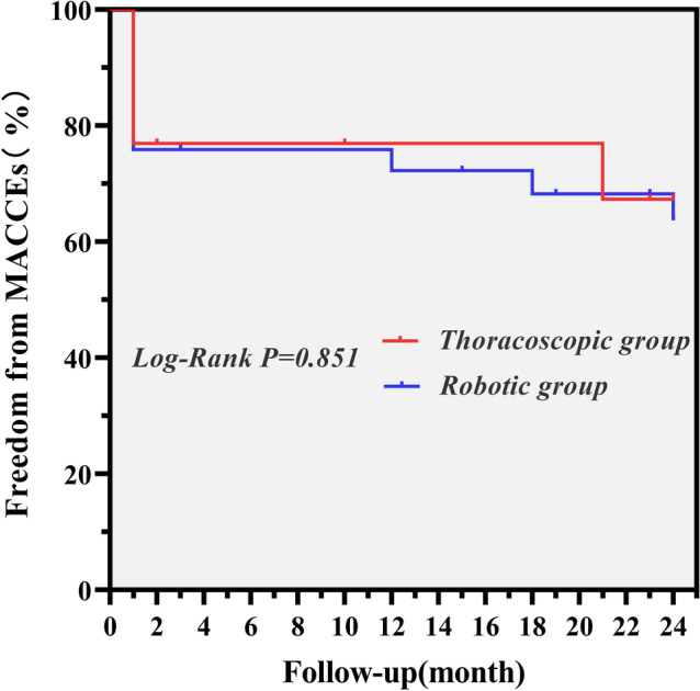
The Kaplan–Meier curve for MACCE
Fig. 3.
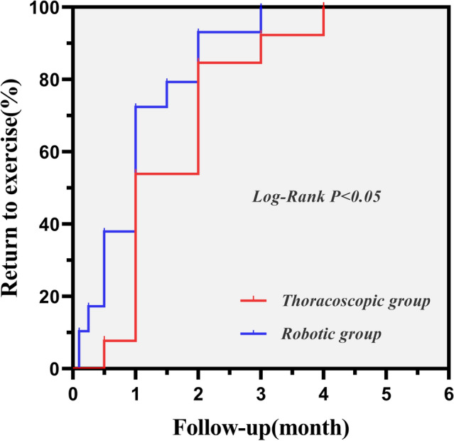
Kaplan–Meier analysis on the rate of returning to exercise
Discussion
Since the first successful robotic cardiac myxoma resection performed by Murphy et al. in 2005 [14], the procedure has been demonstrated as a safe and feasible method with rapid recovery, cosmetic superiority, and improved postoperative quality of life when compared to the conventional sternotomy approach [4, 5, 13, 15–18]. This study aims to explore the differences in clinical outcomes between robotic and thoracoscopic myxoma resection, which are not well documented in previous literature. In our study, both the CPB time and operation time were found to be significantly shorter in the robotic group. We speculated that this difference might result from the fact that the wrist-like articulated instruments of robotic system can move at six degrees of freedom without tremors. In addition, this can help improves the speed and precision of surgical manipulation, thus shortening the CPB and operation time. In comparison, the long-shafted thoracoscopic instruments, to some extent, limits the surgeon’s mobility and can easily cause fatigue. In addition, the process of careful hemostasis is also essential for an uneventful postoperative course. Potential bleeding sites in myxoma resection surgeries include the atriotomy incision, the aortic purse for the cardioplegia needle, and the port incision [4]. Robotic system can provide maximum visualization of bleeding points and intracardiac structure using three-dimension (3D) high-definition imaging [19], which greatly facilitates surgical hemostasis intraoperatively. Significantly, less 24-h postoperative drainage of robotic group was observed in this study, reflecting a better hemostatic effect and less tissue damage. In this study, no conversion to sternotomy was observed in either group, indicating the feasibility of both approaches for myxoma resection.
The current study is the first to compare the postoperative parameters between robotic and thoracoscopic approaches to myxoma resection. In previous studies comparing the robotic and thoracoscopic approaches in mitral valve repair, the ICU duration and postoperative ventilation time were reported to be longer in the robotic group [8]. In contrast, our study found that durations of ICU stay, bed stay, and postoperative mechanical ventilation were significantly lower in the robotic group, which indicates a faster recovery using the robotic technique in myxoma resection. In addition, the robotic group also showed a significantly faster return to exercise after discharge.
Massive allogenic blood transfusion is an independent risk factor for nosocomial pneumonia following cardiac surgeries and plays an important role in postoperative kidney injury [16]. Wei S et al. [8] concluded that thoracoscopic surgeries have a lower transfusion rate than robotic surgeries (52.2% vs. 64.5%) [8]. We found a different trend in the transfusion rates in this study (64.5% for robotic, 66.7% for thoracoscopic); however, no significant difference was observed between the two groups. Besides, there was no significant difference in the total perioperative RBC transfusion between the two groups, whereas the robotic group seemed to receive less intraoperative RBC transfusion.
Cardiac myxoma resection via sternotomy had an early postoperative mortality of 0% to 10% based on previous studies [17, 20–22]. In contrast, no 30-day mortality was observed for either group in this study. This also indicates that both of these two minimally invasive approaches were safe and feasible. Balkhy et al. reported that atrial fibrillation was the most common complication following robotic cardiac surgeries, with an incidence of 12% [23]. Similarly, new-onset atrial fibrillation was also the most frequent complication in this study (19.4% for robotic, 13.3% for thoracoscopic). However, the incidence of atrial fibrillation after robotic surgeries was higher than previously published data [13, 23]. This may be partly due to the limited sample size and a higher proportion of biatriotomy in the robotic group. Although biatriotomy can provide convenience for a large-sized myxoma excision [24], this technique is assumed to be the potential cause of atrial fibrillation after myxoma resection surgery [4, 25]. In this study, up to 25.8% of patients in the robotic group underwent left atrial myxoma resection via biatriotomy, and we speculated that this could be the reason for the high incidence of new-onset atrial fibrillation after robotic surgeries. New-onset stroke rate was low in both study groups (0% for thoracoscopic, 3.2% for robotic). We suggested that the magnified visualization provided by the robotic or thoracoscopic approaches has improved the surgeon’s ability to recognize fragments shed by friable myxoma, which in turn has helped to prevent postoperative stroke. Furthermore, no significant difference was noted between the two groups in terms of in-hospital complications rate and incidence of MACCEs. However, our study has unsurprisingly confirmed that the robotic group involves more costs in comparison with the thoracoscopic group. This particular disadvantage of high costs may limit the application of the robotic technique to some degree. Thus, the thoracoscopic technique could be an effective alternative when the robotic technique is not affordable.
Limitations
First, our study is a non-randomized, retrospective study. We recognize that a selection bias may exist that cannot be fully eliminated. Thus, the lack of randomized and prospective design is an inherent limitation. Second, this study only reported a single-centered experience with limited sample size, which may reduce the power to detect significant differences. A multi-center study with larger sample size and longer follow-up time are still required. Furthermore, the costs cited in our study may not be reproduced in other countries due to the factors such as the inflation rate and different economic levels.
Conclusion
This study demonstrates that both thoracoscopic and robotic approaches are safe and feasible for cardiac myxoma resection. However, regardless of higher expenses, robotic myxoma resection can offer shorter operation-related time, less postoperative thoracic drainage, and faster recovery than total thoracoscopic technique.
Author contribution
Concept—YL, CZ, and SG; design—SG, CZ, and YL; writing—YL; supervision—SG and CZ; data collection—YL, XL, ZL, and CL; analysis—YL and YJ; literature review—YL and YJ; critical review—SG and CZ.
Funding
This study is supported by Natural Science Foundation of Anhui province (1808085MH279).
Data availability
The clinical data used to support the findings of this study are available from the corresponding author upon reasonable request.
Declarations
Conflict of interest
The authors declare that they have no competing interests.
Ethical approval
This is an observational study. The institutional Ethical Review Board of the first affiliated hospital (Anhui Medical University) has confirmed that no ethical approval is required.
Informed consent
Written informed consent about the surgical procedure was obtained from all the patients enrolled in this study.
Consent to publish
Not applicable.
Footnotes
Publisher's Note
Springer Nature remains neutral with regard to jurisdictional claims in published maps and institutional affiliations.
Contributor Information
Chengxin Zhang, Email: zcxayfy@126.com.
Shenglin Ge, Email: ahykdgsl@163.com.
References
- 1.Griborio-Guzman AG, Aseyev OI, Shah H, et al. Cardiac myxomas: clinical presentation, diagnosis and management. Heart. 2022;108(11):827–833. doi: 10.1136/heartjnl-2021-319479. [DOI] [PubMed] [Google Scholar]
- 2.Saadeh AM, Hijazi EM, Saadeh NA. Right atrioventricular myxoma presenting with recurrent syncopal attacks. Am J Case Rep. 2021;22:e927874. doi: 10.12659/AJCR.927874. [DOI] [PMC free article] [PubMed] [Google Scholar]
- 3.Straw S, Sengupta A, Gantenby VK, et al. An enormous right atrial myxoma highlights the role of echocardiography in heart failure pathways: a case report. Eur Heart J Case Rep. 2022;6(2):c42. doi: 10.1093/ehjcr/ytac042. [DOI] [PMC free article] [PubMed] [Google Scholar]
- 4.Kadirogullari E, Onan B, Aydin U, et al. A comparison of robotically-assisted endoscopic versus sternotomy approach for myxoma excision: a single-center experience. Turk Gogus Kalp Damar Cerrahisi Derg. 2020;28(3):450–459. doi: 10.5606/tgkdc.dergisi.2020.19278. [DOI] [PMC free article] [PubMed] [Google Scholar]
- 5.Yang M, Yao M, Wang G, et al. Comparison of postoperative quality of life for patients who undergo atrial myxoma excision with robotically assisted versus conventional surgery. J Thorac Cardiovasc Surg. 2015;150(1):152–157. doi: 10.1016/j.jtcvs.2015.01.056. [DOI] [PubMed] [Google Scholar]
- 6.Cerny S, Oosterlinck W, Onan B, et al. Robotic cardiac surgery in Europe: status 2020. Front Cardiovasc Med. 2021;8:827515. doi: 10.3389/fcvm.2021.827515. [DOI] [PMC free article] [PubMed] [Google Scholar]
- 7.Ma ZS, Yang CY, Dong MF, et al. Totally thoracoscopic closure of ventricular septal defect without a robotically assisted surgical system: a summary of 119 cases. J Thorac Cardiovasc Surg. 2014;147(3):863–867. doi: 10.1016/j.jtcvs.2013.10.065. [DOI] [PubMed] [Google Scholar]
- 8.Wei S, Zhang X, Cui H, et al. Comparison of clinical outcomes between robotic and thoracoscopic mitral valve repair. Cardiovasc Diagn Ther. 2020;10(5):1167–1174. doi: 10.21037/cdt-20-197. [DOI] [PMC free article] [PubMed] [Google Scholar]
- 9.Harky A, Chaplin G, Chan J, et al. The future of open heart surgery in the era of robotic and minimal surgical interventions. Heart Lung Circ. 2020;29(1):49–61. doi: 10.1016/j.hlc.2019.05.170. [DOI] [PubMed] [Google Scholar]
- 10.Kwon HJ, Choi JH, Kim HR, et al. Radiofrequency vs. Cryoballoon vs. Thoracoscopic surgical ablation for atrial fibrillation: a single-center experience. Medicina (Kaunas) 2021;57(10):1023. doi: 10.3390/medicina57101023. [DOI] [PMC free article] [PubMed] [Google Scholar]
- 11.Amabile A, Torregrossa G, Balkhy HH. Robotic-assisted coronary artery bypass grafting: current knowledge and future perspectives. Minerva Cardioangiol. 2020;68(5):497–510. doi: 10.23736/S0026-4725.20.05302-5. [DOI] [PubMed] [Google Scholar]
- 12.Deng L, Zhang GW, Liu ZH, et al. Totally thoracoscopic surgery for atrial myxomas resection and atrial septal defect repair. Eur Rev Med Pharmacol Sci. 2017;21(3):569–575. [PubMed] [Google Scholar]
- 13.Moss E, Halkos ME, Miller JS, et al. Comparison of endoscopic robotic versus sternotomy approach for the resection of left atrial tumors. Innovations (Phila) 2016;11(4):274–277. doi: 10.1097/IMI.0000000000000282. [DOI] [PubMed] [Google Scholar]
- 14.Murphy DA, Miller JS, Langford DA. Robot-assisted endoscopic excision of left atrial myxomas. J Thorac Cardiovasc Surg. 2005;130(2):596–597. doi: 10.1016/j.jtcvs.2004.12.005. [DOI] [PubMed] [Google Scholar]
- 15.Zhao F, Chen T, Tang Y, et al. Totally thoracoscopic surgery for treating left atrial myxoma. Medicine (Baltimore) 2021;100(45):e27819. doi: 10.1097/MD.0000000000027819. [DOI] [PMC free article] [PubMed] [Google Scholar]
- 16.Lu F, Xin Y, Xu JJ, et al. Minimally invasive myxoma resection: a single-center 5 year’s experience. Heart Surg Forum. 2022;25(3):E353–E357. doi: 10.1532/hsf.4689. [DOI] [PubMed] [Google Scholar]
- 17.Schilling J, Engel AM, Hassan M, et al. Robotic excision of atrial myxoma. J Card Surg. 2012;27(4):423–426. doi: 10.1111/j.1540-8191.2012.01478.x. [DOI] [PubMed] [Google Scholar]
- 18.Kesavuori R, Raivio P, Jokinen JJ, et al. Quality of life after robotically assisted atrial myxoma excision. J Robot Surg. 2015;9(3):235–241. doi: 10.1007/s11701-015-0521-4. [DOI] [PubMed] [Google Scholar]
- 19.Yun T, Kim H, Sohn B, et al. Robot-assisted repair of atrial septal defect: a comparison of beating and non-beating heart surgery. J Chest Surg. 2022;55(1):55–60. doi: 10.5090/jcs.21.111. [DOI] [PMC free article] [PubMed] [Google Scholar]
- 20.Pinede L, Duhaut P, Loire R. Clinical presentation of left atrial cardiac myxoma. A series of 112 consecutive cases. Medicine (Baltimore) 2001;80(3):159–172. doi: 10.1097/00005792-200105000-00002. [DOI] [PubMed] [Google Scholar]
- 21.Tasoglu I, Tutun U, Lafci G, et al. Primary cardiac myxomas: clinical experience and surgical results in 67 patients. J Card Surg. 2009;24(3):256–259. doi: 10.1111/j.1540-8191.2008.00797.x. [DOI] [PubMed] [Google Scholar]
- 22.Lukacs L, Lengyel M, Szedo F, et al. Surgical treatment of cardiac myxomas: a 20-year follow-up. Cardiovasc Surg. 1997;5(2):225–228. doi: 10.1016/s0967-2109(96)00084-1. [DOI] [PubMed] [Google Scholar]
- 23.Balkhy HH, Nisivaco S, Torregrossa G, et al. Multi-spectrum robotic cardiac surgery: early outcomes. JTCVS Tech. 2022;13:74–82. doi: 10.1016/j.xjtc.2021.12.018. [DOI] [PMC free article] [PubMed] [Google Scholar]
- 24.Lin Y, Xiao J, Chen J, et al. Treating cardiac myxomas: a 16-year Chinese single-center study. J Cardiovasc Med (Hagerstown) 2016;17(1):44–53. doi: 10.2459/JCM.0000000000000114. [DOI] [PubMed] [Google Scholar]
- 25.Bateman TM, Gray RJ, Raymond MJ, et al. Arrhythmias and conduction disturbances following cardiac operation for the removal of left atrial myxomas. J Thorac Cardiovasc Surg. 1983;86(4):601–607. doi: 10.1016/S0022-5223(19)39129-9. [DOI] [PubMed] [Google Scholar]
Associated Data
This section collects any data citations, data availability statements, or supplementary materials included in this article.
Data Availability Statement
The clinical data used to support the findings of this study are available from the corresponding author upon reasonable request.