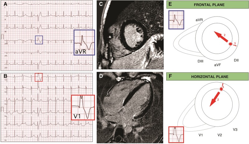
Figure 3.
Representative case of a patient with frequent PVCs with RBBB morphology SA-IntA and absence of LGE on CMR. A 16-year-old athlete with no family history of heart diseases and no symptoms. Echocardiography was normal. A 12-lead ECG showed isolated premature ventricular beats with a right bundle branch block/superior axis morphology and distinguished by the presence of a qR pattern in both leads aVR and V1 (A, B). Cardiac magnetic resonance, short axis (C) and 2-chamber long axis (D) views demonstrated absence of LGE. Depolarization forces in frontal and transversal plane of PVCs RBBB/SA-IntA morphology. The main depolarization vector is directed away from its origin point. If PVCs originate from LV inferior and infero-lateral endocardial wall an initial depolarizing force is directed away from leads V1 and aVR, in an endo-epicardial activation sequence, which gives rise to a q wave (E, F, arrow 1). The volume arrow points in the main depolarization vector’s direction (E, F, arrow 2).

