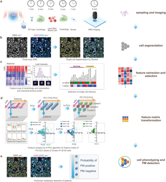
Figure 1.

Workflow of stimulated Raman molecular cytology (SRMC). a) Sample preparation and SRS imaging. b) Single cell segmentation based on three‐color SRS images. c) Single cell feature extraction and selection. d) Feature matrix transformation from raw features by K‐PCA algorithm. e) Cell phenotyping by K‐PCA, and PM positive and negative detection by machine learning classifiers.
