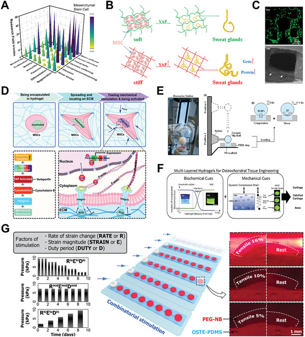
Figure 4.

Mechanical stimulation on mesenchymal stem cell (MSC) behaviors by hydrogel‐based platforms. A) 3D bar graph of mechanical stimulation on MSC for various cell activities and biomedical applications. The research conditions are evaluated based on h‐index of each field calculated by paper publication and citation data from Scopus. B) Schematic illustration of bioprinted stiffer hydrogels that can upregulate Yes‐associated protein (YAP) level and heighten the expression of sweat gland cell phenotype. Reproduced with permission.[ 178 ] Copyright 2020, Elsevier. C) Fluorescent and SEM images of MSC (green fluorescence or white arrows) spreading and growing in macroporous silk‐gelatin 3D hydrogel scaffold. Reproduced with permission.[ 122 ] Copyright 2021, Elsevier. D) Schematic illustration of mechanical stimulation from dynamic stiffening matrix. The stiffness stress is transmitted via cell cytoskeleton and induces YAP nuclear localization and ultimately potentiates paracrine response. Reproduced with permission.[ 82 ] Copyright 2020, the Royal Society of Chemistry. E) Schematic illustration of bioreactor operation for compression and shear forces to MSC‐seeded hydrogel. Reproduced with permission.[ 195 ] Copyright 2017, Springer Nature. F) Multilayer hydrogel with specific biochemical cues, matrix stiffness, and dynamic mechanical loading for osteochondral tissue engineering. Reproduced with permission.[ 197 ] Copyright 2015, Elsevier. G) Illustration and images of combinatorial mechanical stimulation patterns in combination with defined levels of rate of strain change, strain magnitude, and duty period. Reproduced with permission.[ 190 ] Copyright 2021, American Association for the Advancement of Science.
