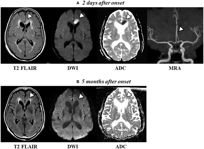
Figure 1.
(A) Axial T2 FLAIR scan and diffusion-weighted imaging (DWI) revealed a high-intensity signal [apparent diffusion coefficient (ADC) shows a low signal intensity, indicating acute cerebral ischemia] in the left frontal lobe and midbody of the CC 2 days after onset. Magnetic resonance angiography (MRA) images revealed a vessel occlusion of the left A2. (B) Axial T2 FLAIR and DWI show a change to a low-intensity signal (conversely, ADC shows a high-intensity signal, indicating a chronic phase) in the left frontal lobe and the midbody of the CC 5 months after onset.

