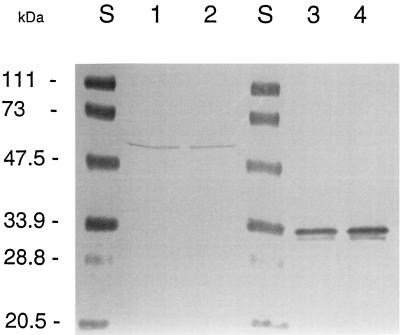
FIG. 7.
Western blotting of BusAB and OpuAC proteins. Proteins were separated by SDS-PAGE (12.5% polyacrylamide) and transferred to a nitrocellulose membrane. Shown are proteins of the membrane fractions of L. lactis cells grown in M17 (lanes 1) or in M17 containing 0.3 M NaCl (lane 2) and proteins from whole-cell extracts of B. subtilis JH642 obtained after growth on LB (lane 4) or LB containing 0.5 M NaCl (lane 5). BusAB and OpuAC were detected with an antiserum against the purified OpuAC protein of B. subtilis JH642. The molecular mass marker was from Bio-Rad (lanes S).

