Figure 2.

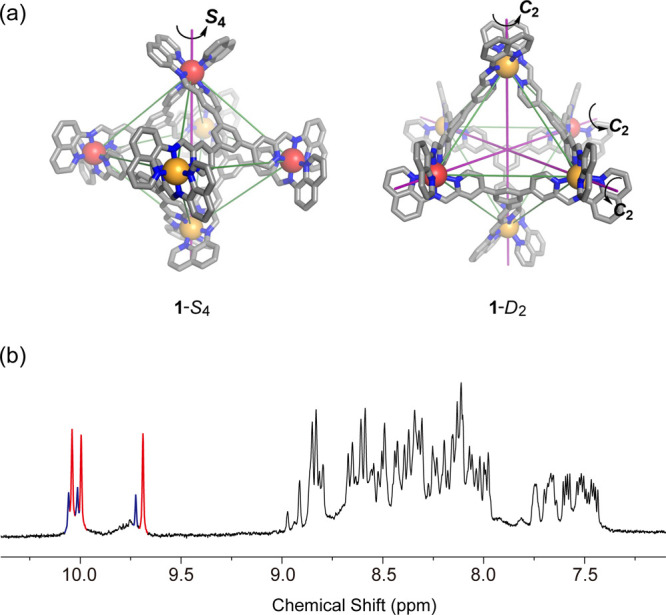
(a) PM7-optimized structure of the S4 and D2 diastereomers of cage 1. Color scheme: Δ-Zn, yellow; Λ-Zn red. (b) 1H NMR spectrum (400 MHz, 298 K, CD3CN) of cage 1. The two sets of imine peaks with a 1:1:1 integration ratio are highlighted.

(a) PM7-optimized structure of the S4 and D2 diastereomers of cage 1. Color scheme: Δ-Zn, yellow; Λ-Zn red. (b) 1H NMR spectrum (400 MHz, 298 K, CD3CN) of cage 1. The two sets of imine peaks with a 1:1:1 integration ratio are highlighted.