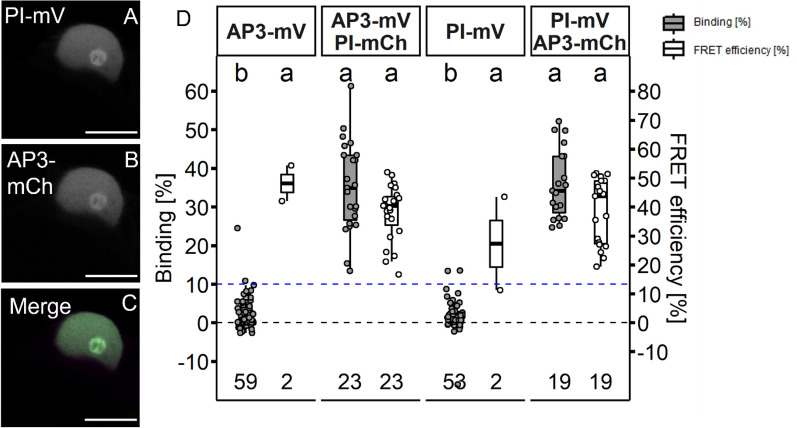
Fig. 7.
AP3 and PI homomerization in N. benthamiana leaf cells. AP3 and PI fused to the indicated FPs were expressed via the UBQ10 promoter and imaged three days after infiltration. A–C Co-localisation of PI-mV and AP3-mCh in N. benthamiana leaf cells (A PI-mV signal. B AP3-mV signal. C Merged signal). Co-expression of AP3 with PI lead to an accumulation of both AP3 and PI in the nucleolus (for individual expressed AP3 and PI compare Fig. 3; Scalebars: A–C 10 µm). D BINDING [%] (grey) and FRET efficiencies [%] (white) for AP3-mV, AP3-mV PI-mCh, PI-mV and PI-mV AP3-mCh. Analysis was done as described in Fig. 5. Average BINDING between AP3 and PI was high in both measured directions (35.25% ± 11.65 for AP3/PI and 35.97% ± 8.99 for PI/AP3) with mean FRET Efficiencies of ~ 38%. Statistical groups were assigned after multiple comparison with Kruskal–Wallis and a Post hoc test using the criterium Fisher’s least significant difference (alpha parameter is 0.05) (Dashed blue line marks the BINDING cut-off of 10%; Number of repetitions are indicated below BINDING values and number of images with BINDING above 10% are indicated below the FRET efficiency values in the bottom of the plot)

