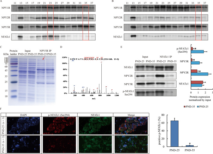
Fig. 3.
Special protein complex of NPY2R/NPY5R/NFATc1 in SG before puberty onset. A, B Gel filtration assay show the distribution of NPY receptors and NFATc1 in VS of PND-25 (A) and PND-35 (B). Red frames highlighted the fraction of potential target protein peak. C Coomassie brilliant blue staining shows the proteome interacted with NPY5R in VS of PND-25 and PND-35. Red arrow highlights a specific band at 140 kDa only appears in PND-25. D Original secondary mass spectrum shows the most confident protein (NFATc1) identified by mass spectrum. E Western blot confirm the interaction between NFATc1 and NPY2R/NPY5R via phosphorylated form. F IF show the localization of NFATc1 and p-NFATc1 at Ser294 in VS of PND-25 and PND-35 (magnification ×200). The SG specimens from two individuals were used for gel filtration, immunoprecipitation and western blot. The SG specimens from three individuals were used for immunofluorescence. Statistical analysis and abbreviated description can be referred to Fig. 1 legend

