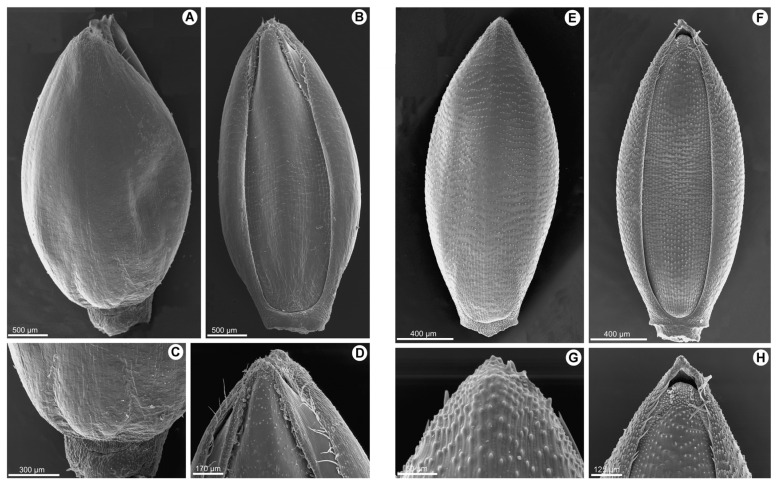
Figure 3.
(A–D). Scanning electron micrographs of the upper anthecium of Panicum deustum: (A). dorsal view; (B). ventral view; (C). base of the upper lemma; and (D). apex of the upper palea. (E–H). Scanning electron micrographs of the upper anthecium of Panicum trichocladum: (E). dorsal view; (F). ventral view; (G). apex of the upper lemma; and (H). apex of the upper palea [vouchers: (A–D): Bequaert 3398; (E–H). Hitchcock 24927].

