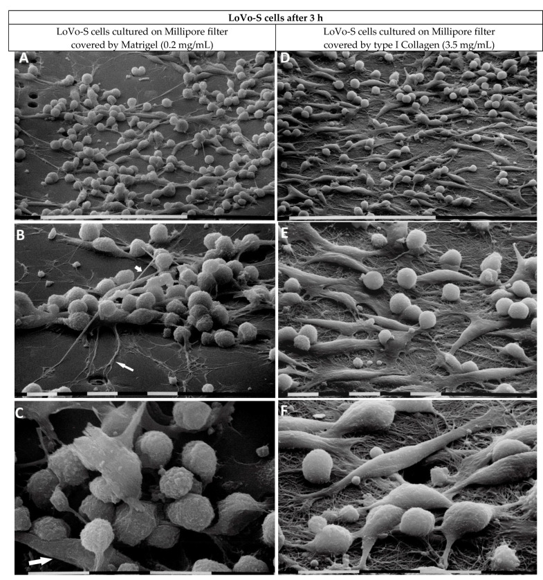
Figure 4.
LoVo-S cells cultivated for 3 h on Millipore filter with 8 µm pores covered by Matrigel (0.2 µg/µL) mimicking the BM or type I Collagen (3.5 µg/µL) mimicking the collagen arrangement of the desmoplastic lamina propria. LoVo-S cells cultured on Matrigel (0.2 mg/mL) cover most of the Millipore pores. The cells appear grouped, and all of them show tight cell–cell contacts. Bar = 100 µm (A). Long filopodia (long arrow) for exploring the microenvironment, as well as intercellular tunneling nanotubes (short arrow) for cell interplay, are visible. Bar = 10 µm (B). The cells show a globular shape with exosomes and microvesicles on their cytoplasmic surface, but very few elongated-mesenchymal-shaped cells are also present (arrow). Bar = 10 µm (C). LoVo-S cells cultivated on highly concentrated type I Collagen (3.5 mg/mL) appear less grouped than the same cultivated on Matrigel. Bar = 100 µm (D). Both elongated-mesenchymal phenotypes, developing filopodia or lamellipodia, and globular-shaped cells are visible. Bar = 10 µm (E). The cells firmly adhere to the collagen fibril surface and most of them appear very smooth with no extravesicles. Single fibrils form a collagen meshwork with small inter fibrillar spaces or pores. Bar = 10 µm (F).

