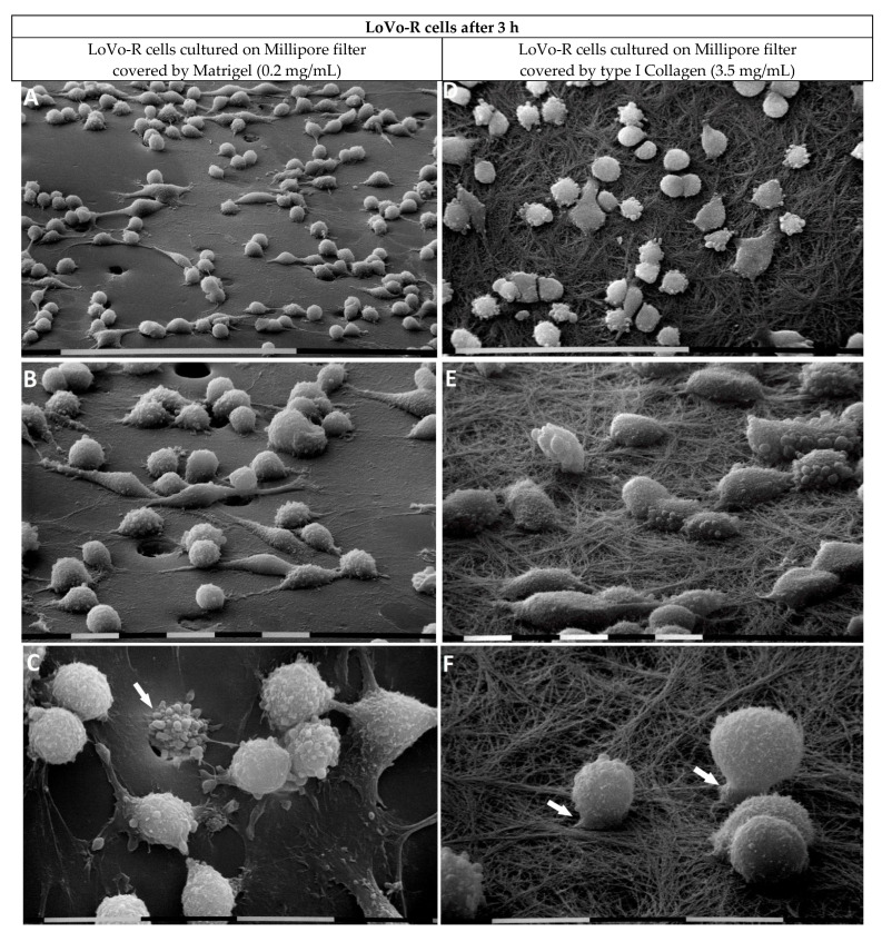
Figure 5.
LoVo-R cells cultivated for 3 h on Millipore filter with 8 µm pores covered by Matrigel (0.2 µg/µL) mimicking the BM or type I Collagen (3.5 µg/µL) reproducing the collagen meshwork of a desmoplastic lamina propria. LoVo-R cells growing on Matrigel are more isolated than the LoVo-S cells (see Figure 3A). Bar = 100 µm (A). LoVo-R cells primarily show a globular shape with protruding exosomes and microvesicles on the surface, but few isolated elongated-fusiform mesenchymal-shaped cells are detectable. Bar = 10 µm (B). Globular-shaped cells developing extravesicles. A cell passing through a Millipore pore (arrow). Bar = 10 µm (C). LoVo-R cells cultivated on type I Collagen meshwork are isolated and do not show cell–cell contact. Bar = 100 µm (D). All the cells, adhering to the collagen fibrils, exhibit a rounded or globular shape and develop exosomes and microvesicles. Bar = 10 µm (E). Isolated globular-shaped cells develop cytoplasmic protrusions or short filopodia, which penetrate into the collagen layer (arrows). Bar = 10 µm (F).

