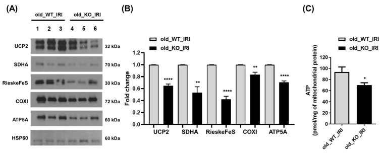
Figure 4.
Mitochondrial proteins decreased in NRF2 KO mice during the recovery phase of IRI. Twelve-month-old WT and NRF2 KO mice underwent IRI to induce AKI. After 4 weeks of injury, mitochondria were extracted from kidney tissues using a mitochondria isolation kit. The mitochondrial fractions were assessed via Western blotting to determine the levels of mitochondrial proteins, including UCP2, SDHA, RieskeFeS, COXI, ATP5A, and HSP60. HSP60 was used as a mitochondrial loading control. Representative blots are shown on the left (A), and densitometric images of the blots are shown on the right (B). (C) Measurement of the ATP content was performed using an ATP assay kit to evaluate the amount of mitochondrial ATP in the kidney mitochondrial fraction. * p < 0.05, ** p < 0.01, **** p < 0.0001 vs. old_WT_IRI group. Abbreviations: IRI, ischemia-reperfusion injury; NRF2, nuclear factor erythroid-2-related factor 2; KO, knockout; AKI, acute kidney injury; UCP2, uncoupling protein-2; SDHA, succinate dehydrogenase complex flavoprotein subunit A; COXI, cytochrome c oxidase subunit 1; ATP5A, ATP synthase F1 subunit alpha; ATP, adenosine triphosphate.

