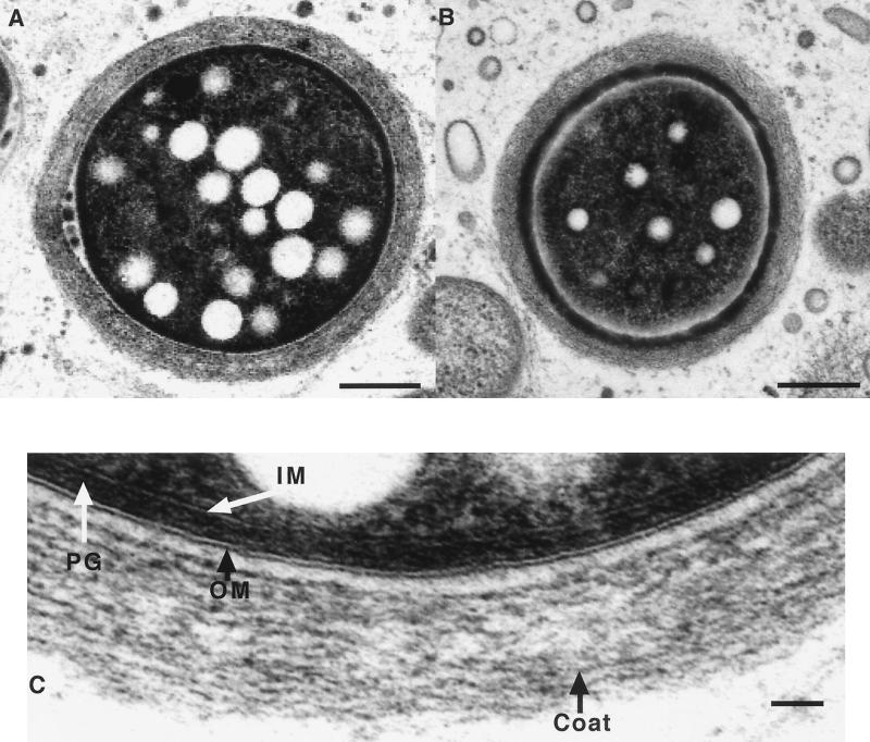
FIG. 9.
Transmission electron microscopy of DK2657 (csgA) spores. One-week-old fruiting body spores from CF plates containing an inducer of β-lactamase were harvested and prepared for transmission electron microscopy as described in Materials and Methods. (A and B) Low-magnification images of two different spores; sections are not necessarily medial, and the apparent difference in size of the spores is related to the position of the section in the spore. Bar = 1 μm. (C) Enlargement of the wall of the cell in panel A. IM, inner membrane; PG, peptidoglycan; OM, outer membrane. Bar = 0.1 μm.

