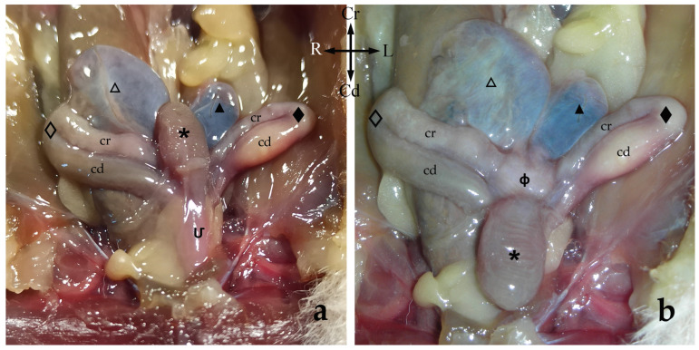
Figure 2.
Ventral view of the female sugar glider’s apparatus urogenitalis in situ (a) and with the urinary bladder displaced caudally (b). Fresh specimen: (Մ) sinus urogenitalis; (ф) sinus vaginalis; (🞳) urinary bladder. Right-side structures are shown with empty patterns whereas left-side structures are marked with filled patterns: (◇, ◆) lateral vaginae with cranial loop (cr) and caudal loop (cd); (△, ▲) uteri.

