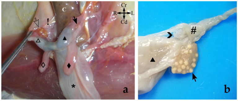
Figure 4.
(a) Ventrolateral view of the sugar glider’s apparatus urogenitalis in situ. Fresh specimen: The urinary bladder is reflected caudally to expose the uterii and vaginae. The (△) right uterus has been displaced ventrally to display the right ovary and its peritoneal folds: (⇨) right ovary; (!) suspensory ligament of the ovary; (🞳) urinary bladder. Right structures are represented by an empty pattern and left structures with a filled pattern: (◇, ◆) lateral vaginae; (△, ▲) uteri; (➞) left ovary; (C) colon. (b) Dorsal view of the female sugar glider’s apparatus genitalis ex situ: ovary and uterine tube. Specimen is fixed in formalin. The oviduct is included in a pouch formed by the peritoneal folds (mesovarium and mesosalpinx); (▲) left uterus; (>) uterine tube; (#) pouch; (➞) left ovary.

