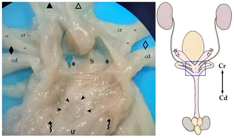
Figure 6.
On the right, a general sketch of the female sugar glider urogenital system ex situ (dorsal view) with the area’s detailed photograph. On the left, view of the floor of the cranial part of the (Մ) urogenital sinus that has been opened by cutting its roof in the midline and moving each half laterally. The vaginal sinus (ф) has also been opened dorsally. In this specimen, the central vagina (marked with a discontinuous oval shape) connects the vaginal and urogenital sinuses. Specimen is fixed in formalin. (⇝) Outlet orifices of the lateral vaginae; (➤) external urethral orifice; (“) ureters; (S) septum. Right structures are represented by an empty pattern and left structures with a filled pattern: (△, ▲) bodies of the uteri; (◇, ◆) lateral vaginae with cranial loop (cr) and caudal loop (cd).

