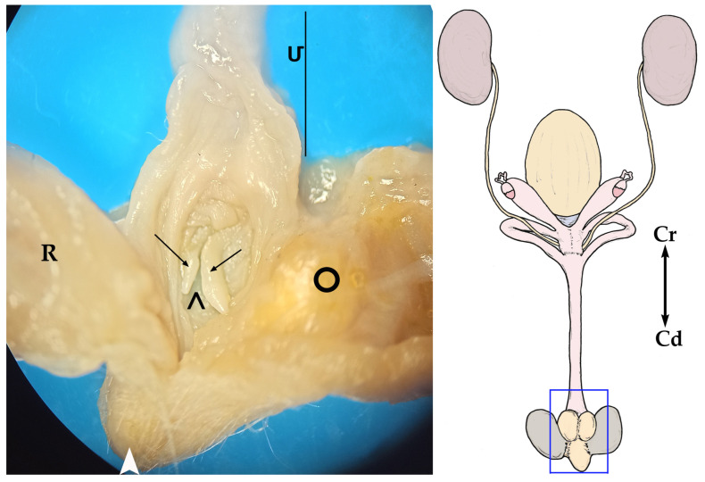
Figure 8.
On the right, a general sketch of the female sugar glider urogenital system ex situ, (dorsal view) with the area’s detailed photograph. On the left, magnified image of the cloacal area; view of the floor of the caudal part of the urogenital sinus (Մ). The sinus has been opened by cutting its roof in the midline and moving each half laterally. (R) Rectum; ( ) cloacal orifice; (➝) clitoris; (>) fossa clitoridis; (○) right paracloacal gland.

