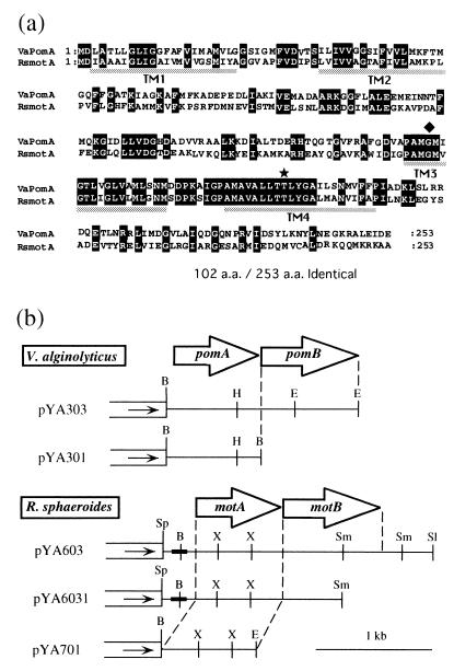
FIG. 1.
(a) Amino acid alignments of V. alginolyticus PomA and R. sphaeroides MotA. White letters in black boxes, diamond, and star show identical residues, the mutation site of the PomA mutant VIO586 (G154R), and the threonine residue that is highly conserved among all species of MotA, respectively. Dotted bars indicate putative transmembrane regions. (b) Restriction map of plasmids. White boxes and the direction of the arrowheads indicate the vector part of pSU41 and the direction of translation from the lac promoter, respectively. Insertional fragments are indicated by solid lines. The bold lines in the inserted fragments indicate the region of the native promoter. The open arrows show the coding regions of PomA, PomB, MotA, and MotB. Abbreviations: B, BamHI; H, HindIII; E, EcoRI; Sp, SphI; X, XhoI; Sm, SmaI; Sl, SalI.

