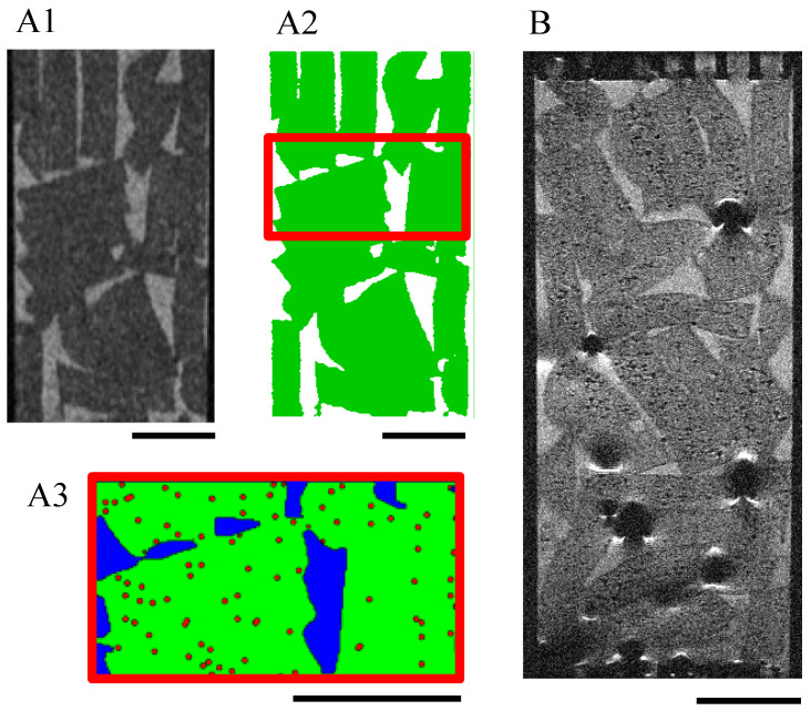
Figure 9.
MRI acquisitions of a scaffold stack within a perfusion bioreactor. T2-turbo-RARE sequences were performed on slices of 250-μm thickness and 55-μm resolution. (A1–A3): Bioreactor without cells: the grayscale images (A1) were segmented into the fluid (white) and the hydrogel scaffolds (green) (A2). The volume of interest (6.55 mm high) defined for the LBM simulations is delimited by red lines. The hydrogel scaffolds (green) of this volume were numerically seeded with spheroids (red dots) of 135 μm diameter (A3). The black scale bars stand for 4 mm. (B): Initial seeding was 100,000 cells per scaffold; the cells were pre-labeled with SPIONLA-HSA, 20 μg[Fe] cm−2. Cells were fixed with PFA 4% after culturing for 18 days in dynamic conditions (10 mL min−1). Cell volume estimation is reported on the calibration curve in Figure 7D. The scale bar stands for 4 mm.

