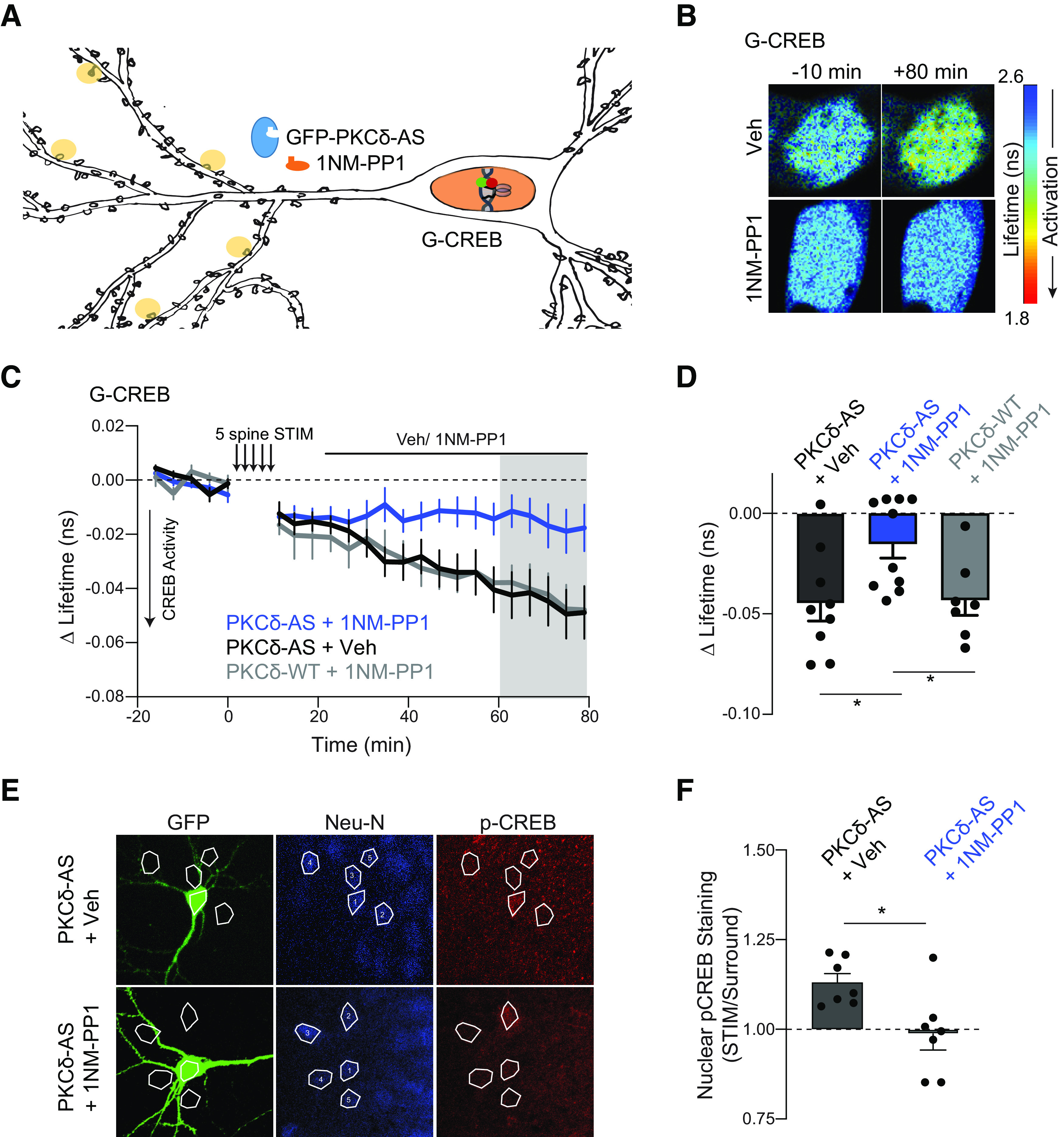
Figure 9.

Long-lasting PKCδ activity regulates CREB activity. A, Schematic of five spine stimulation of CA1 neurons in which PKCδ was replaced by PKCδ-AS. CREB activity was monitored in the nucleus using a CREB sensor. B-D, Representative lifetime images (B), time course (C), and quantification (D) of mean lifetime change of CREB sensor in the nucleus before and after 5 spine stimulation and vehicle (n = 9 neurons) or 1NM-PP1 (1 μm, n = 10 neurons) application 10 min after stimulation to inhibit long-lasting PKCδ activity. Gray shading represents the time of quantification in D. *p = 0.016 (two-tailed unpaired t test). E, Representative images and ROIs of p-CREB signal in nuclei of neurons expressing GFP-PKCδ-AS (GFP) and surrounding neurons. Neurons were simulated with 5 spine stimulation, after which vehicle (n = 7 neurons) or 1NM-PP1 (1 μm, n = 7 neurons) was applied. Neurons were fixed 60 min after stimulation and processed for immunostaining. F, Quantification of p-CREB fluorescence signal in nuclei of stimulated neurons (ROI 1) normalized to signal in nuclei of surrounding neurons (average ROI 2-5, one sample t test of normalized p-CREB signal compared with 1, Veh: p = 0.002, NMPP1 ns p = 0.78). *p = 0.016 (between-group comparison of unpaired t test).
