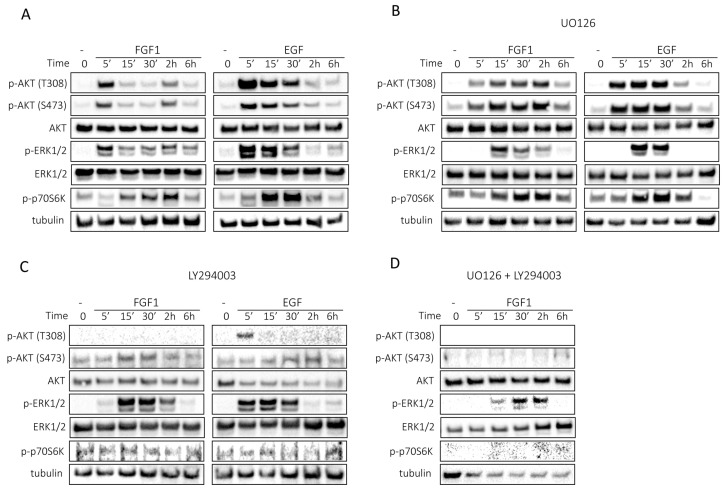
Figure 3.
Kinetics of FGF1- or EGF-stimulated cell signaling in MCF-7 cells. Serum-starved MCF-7 cells were treated with 10 ng/mL FGF1 or EGF in the absence (A) or presence of MEK inhibitor (20 µM UO126) (B) or presence of PI3K inhibitor (20 µM LY294002) (C) or a mixture of MEK inhibitor (20 µM UO126) and PI3K inhibitor (20 µM LY294002) (D) for different times: 0, 5 min, 15 min, 30 min, 2 h and 6 h. Subsequently, cell lysates were subjected to Western blotting with specific antibodies to assess the activation of cellular signaling pathways, including AKT/mTOR and ERKs.

