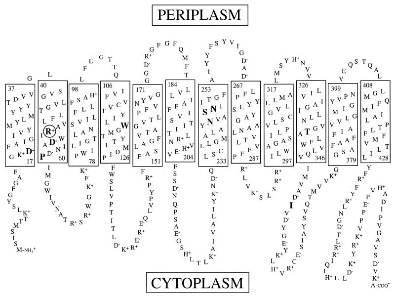
FIG. 1.
Toplogical model of the melibiose carrier. Rectangles represent transmembrane domains. The numbers at the top and bottom of each rectangle indicate the first and last residue of each helix. The circled residue in helix II was subjected to site-directed mutagenesis in this study. Large bold residues indicate the positions of isolated second-site revertants. The model is based on that described by Pourcher et al. (34).

