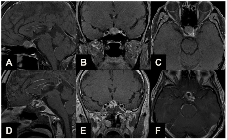
Figure 3.
Illustrative case of a Type 2 sec. Barazi PA suitable for ETTA. (A–C) Midsagittal (A), coronal (B) and axial (C) pre-operative contrast-enhanced T1-weighted MR images of a 63-year-old male patient previously treated with a standard endoscopic endonasal approach for a non-functioning pituitary macroadenoma. Years later, a linearly progressing supradiaphragmatic recurrence with sub-frontal extension was observed. He underwent ETTA, which achieved near-radical resection (a small remnant was revealed with post-operative imaging posteriorly) with an unremarkable clinical course. (D–F) Midsagittal (D), coronal (E) and axial (F) post-operative contrast-enhanced T1-weighted MR images.

