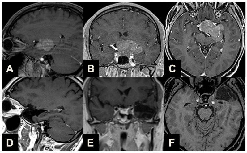
Figure 5.
Illustrative case of a large PA suitable for combined ETTA–TCA. (A–C) Parasagittal (A), coronal (B) and axial (C) pre-operative contrast-enhanced T1-weighted MR images of a 55-year-old male patient complaining of visual disturbances, with a clinically manifest bitemporal hemianopsia and left eye visual impairment consistent with second left cranial nerve involvement. Imaging and laboratory exams reported a non-functioning macroadenoma with a significant left lateral extension, invading and obliterating the ipsilateral basal cisterns. He underwent ETTA with a left TCA in the same surgical session, which achieved near-radical resection. Two millimetric remnants were revealed with post-operative imaging at the level of left cavernous sinus and interpeduncular cistern. The patient experienced a clinically silent left temporal pole ischemia, severe post-operative panhypopituitarism and diabetes insipidus, which required persistent complete substitution therapy; conversely, a complete resolution of pre-operative visual acuity and field deficits was observed. (D–F) Parasagittal (D), coronal (E) and axial (F) post-operative contrast-enhanced T1-weighted MR images.

