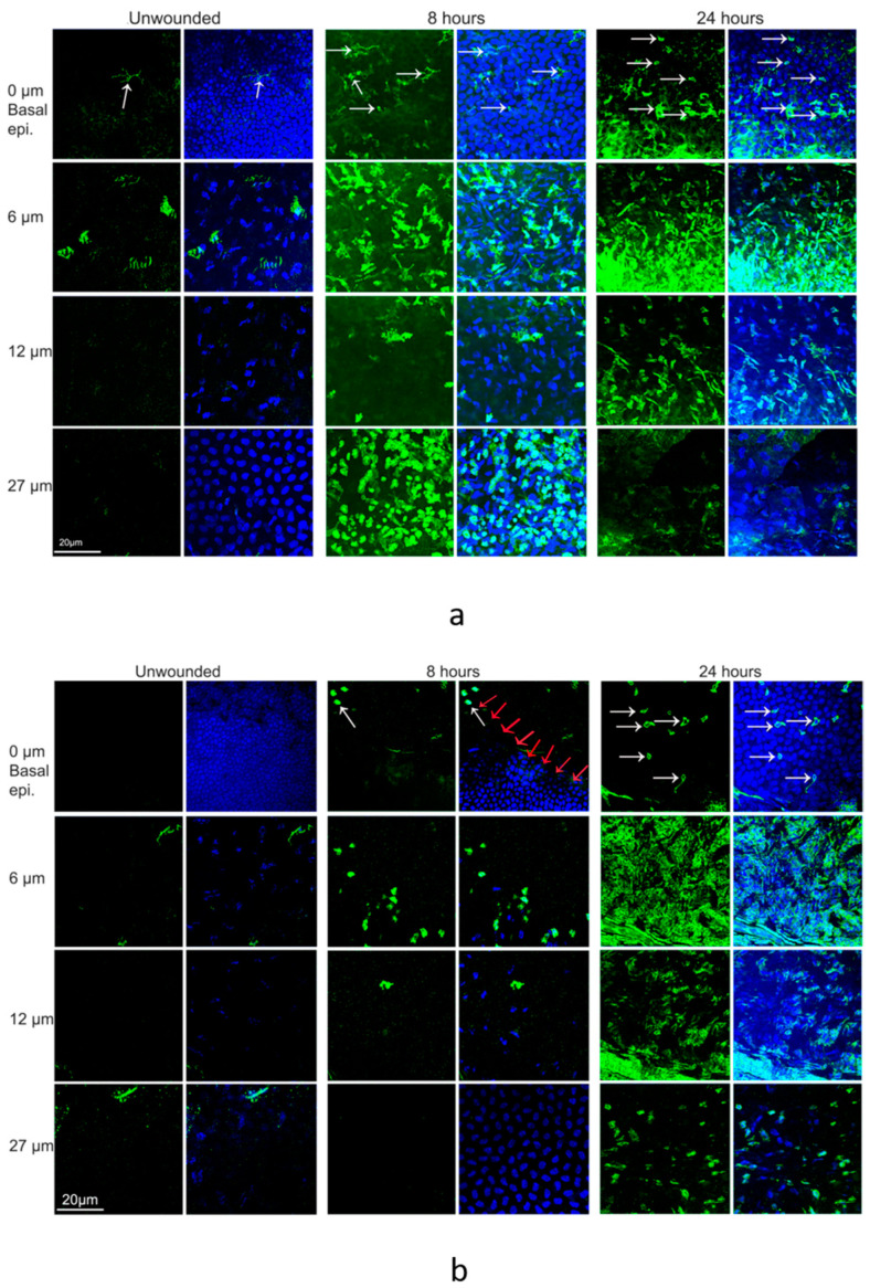
Figure 5.
Distribution of CD45+ cells in unwounded and wounded normoglycemic mice at 8 h and 24 h. Corneas from wounded mice were harvested at 8 h and 24 h for immunohistochemistry. Corneas were immunostained with an anti-CD45 antibody (green) and DAPI (blue). (a) Representative images illustrating the distribution of CD45+ cells from the basal epithelia to endothelium in the peripheral cornea. Depth of the corneal section is shown on the left. (b) Distribution of CD45+ cells from the basal epithelia to endothelium in the central cornea (red arrows point to the wound edge, and white arrows point to CD45+ cells in the sub-basal epithelium).

