FIG. 3.
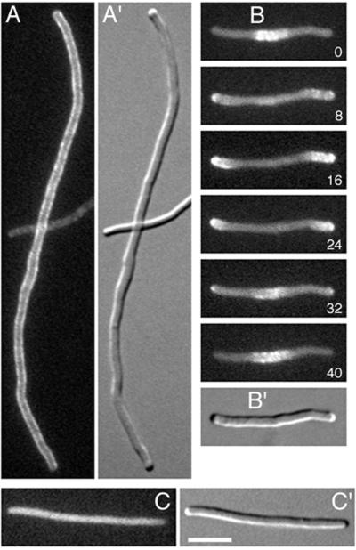
Gfp-MinC localization in filaments. Fluorescence (A to C) and differential interference contrast (A′ to C′) images showing the distribution of Gfp-MinC in cells in which FtsZ ring assembly is blocked. Cells were grown at 37°C with 50 μM IPTG. (A) Gfp-MinC localization in MinE− filaments of strain PB114(λDR155)/pDR175 [ΔminCDE(Plac::minD)/λpR::gfp-minC]. (B) Time-lapse images of Gfp-MinC localization in a SfiA-induced filament of strain PB103(λDR144)/pDR175 [WT(Plac::sfiA)/λpR::gfp-minC]. Times are indicated in seconds. (C) Random distribution of Gfp-MinC in a SfiA-induced filament of strain PB114(λDR144)/pDR175 [ΔminCDE(Plac::sfiA)/λpR::gfp-minC]. Bar, 5 μm.

