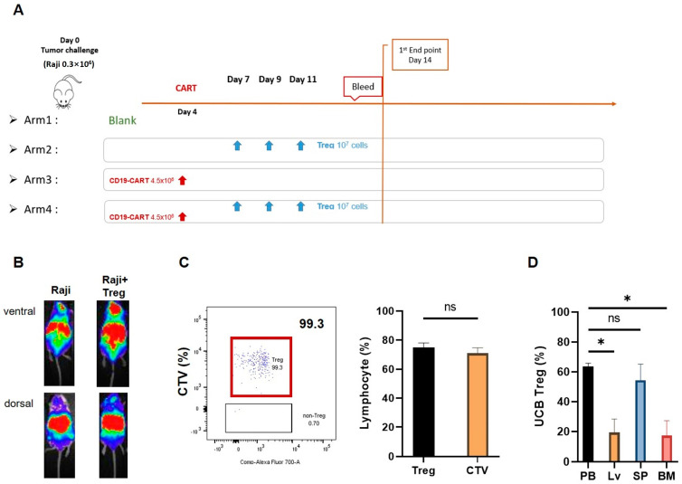
Figure 2.
UCB Treg cells show persistence in a xenogeneic lymphoma model. (A) Xenogeneic lymphoma model and UCB Treg injection. Female Rag2−γc−mice were injected with 0.3 × 10⁶ RFP−fLuc−Raji cells by tail vein on Day 0. On day 4, after mice displayed engraftment of the tumor mass as per in vivo imaging, a one-time tail vein injection of 4.5 × 10⁶ CD19 CAR-T cells was administered. on day 4, followed by 1 × 10⁷ UCB-Treg cells administration by tail vein injection on days +7, +9, and +11. Peripheral blood and mouse organ tissue samples were harvested from day 14 onward. (B) Non-invasive bioluminescence detects tumors in UCB Treg recipients. Single tail vein injection of 0.3 × 10⁶ Raji cells led to the development of disease in NSG mice +/− UCB Treg cells, where tumor engraftment is higher in the areas corresponding to the liver and brain in control mice compared to differential preference for the spleen and PB in the +UCB Treg arm. (C) Representative flow cytometric plots show that almost all the circulating non-tumor cell population in Treg recipients was comprised of CTV-labeled cells, and all CTV labeled cells demonstrated the phenotype of Treg cells (CD4+CD25+), p = n.s., student t-test. CTV, CellTraceTM Violet; PB, peripheral blood. (D) UCB Tregs concentrate in the PB and liver in the xenogeneic lymphoma model. Flow analysis was performed in the harvested organ cell suspension upon euthanasia performed on day 14. The PB and spleen have a higher UCB Treg distribution. Error bars represent SEM (n = 7); statistical differences compared with PB were quantified by one-way ANOVA using GraphPad Prism software (version 9.5.0): * p < 0.05.

