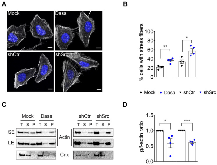
Figure 2.
Src kinase controls actomyosin cytoskeleton organization in HeLa cells. (A) Confocal microscopy images of control (mock and shCtr) and Src-impaired (Dasatinib-treated and shSrc) HeLa cells stained with phalloidin for f-actin (greyscale) and with DAPI (blue). Scale bar, 10 µm. (B) Percentage of cells with stress fibers quantified from images similar to those shown in (A). Values are the means ± SEM (n = 4); p-values were calculated using a two-tailed unpaired Student’s t-test, * p < 0.05, ** p < 0.01. (C) Immunoblots showing the levels of globular (g)- and filamentous (f)-actin in HeLa cells treated as in (A). g- and f-actin from the total cell lysates (T) were separated by ultracentrifugation. g-actin was recovered from supernatant fractions (S) while f-actin was associated with pellet fractions (P). Calnexin was used as a loading control (Cnx). Actin levels are shown with both short exposure (SE) and long exposure (LE). (D) Quantification of the g-/f-actin ratio was conducted using immunoblot signals. Values are the means ± SEM (n = 4); p-values were calculated using a two-tailed unpaired Student’s t-test, * p < 0.05, *** p < 0.001. In (B,D), each dot or triangle corresponds to an independent experiment.

