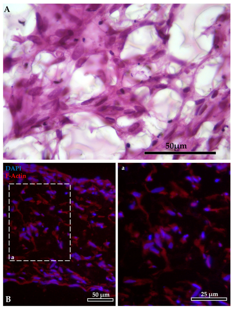
Figure 3.
Intricate mesh generated by the fibroblasts inside the AlvetexTM scaffold: (A) staining with HE. Fibroblast organization inside the AlvetexTM scaffold: (B) F-actin stained in red with phalloidin-iFluor 594 reagent; nuclei counterstained in blue with DAPI (panel a shows F-actin staining at higher magnification).

