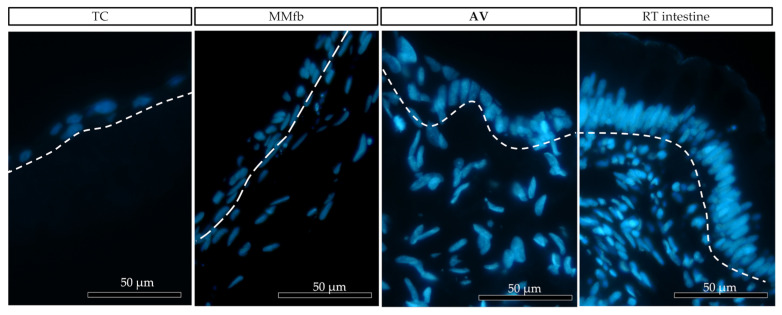
Figure 9.
Representative images of the organization of epithelial (above the dotted line) and fibroblast cell (below the dotted line) growth on the different platforms: ThinCert® (TC) inserts, Matrigel Matrix® enriched with fibroblasts (MMfb), Alvetex™ scaffold (AV), and the RT intestine. Nuclei were stained with DAPI (in blue). The three platforms showed an increasing similarity (from left to right) with their in vivo counterpart.

