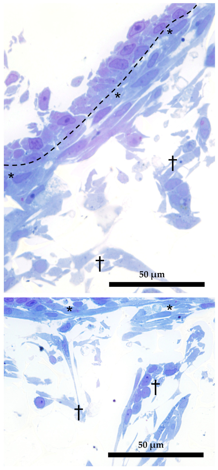
Figure 11.
Dual organization of RT fibroblasts within the AV scaffolding (semithin section stained with toluidine blue). Fibroblasts in the close proximity of the epithelial cells assumed a thin and elongated morphology (asterisks); those located more in-depth acquired a loose distribution and a varied phenotype (crosses). Dotted line underlies the boundary between epithelial and fibroblasts cells.

