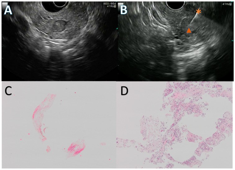
Figure 2.
Endoscopic ultrasound guided fine needle biopsy (EUS FNB) of a pancreatic neuroendocrine tumor. Solid, 10 mm, hypoechogenic round lesion in the pancreatic head (A) first punctured with a 20 G Procore needle with inconclusive results, then with a 22 G Acquire needle (B) during a repeat EUS FNB. The first EUS FNB showed only fibrin and red blood cells (C), while the second one retrieved multiple neuroendocrine-like cells (D). Figure (C,D) are at ×4 magnification. The diagnosis was confirmed by a strong anti-synaptophysin antibody staining on immunohistochemistry. *: 22 G Acquire needle, ▲: neuroendocrine tumor.

