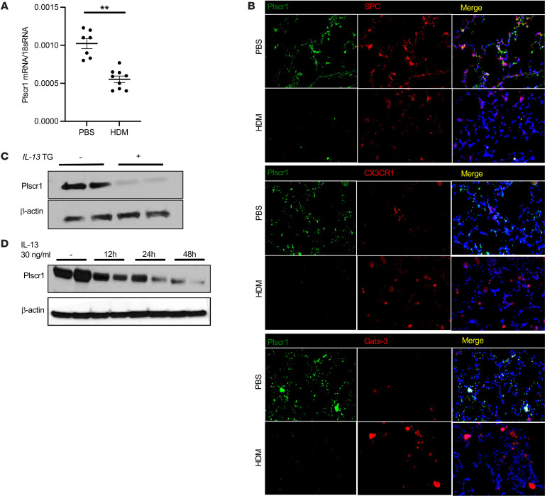
Figure 1. The expression of Plscr1 is inhibited by IL-13 and Type 2 inflammation.
(A) WT mice were subjected to HDM administration, lung Plscr1 mRNA expression was assessed by quantitative real-time PCR. Values are mean± SEM with 7–9 mice in each group. Data was assessed with unpaired Student’s t-test. **P ≤ 0.01. (B) Lungs from WT mice with or without HDM challenges were sectioned, SPC, CX3CR1, or Gata-3 was labeled with red fluorescence (Alexa Fluor 594) and Plscr1 was labeled with green fluorescence (Alexa Fluor 488). Nuclei are stained with DAPI (blue). Images are representative of 3 mice. (C) Whole lung lysates from WT (IL-13 Tg (–)) and IL-13 Tg (+) mice were isolated and Plscr1 protein level was then evaluated by Western immunoblot analysis as noted. (D) BAL inflammatory cells were isolated from WT mice. Cells were treated with 30 ng/mL IL-13, and Plscr1 protein level was evaluated using Western immunoblot analysis as noted.

