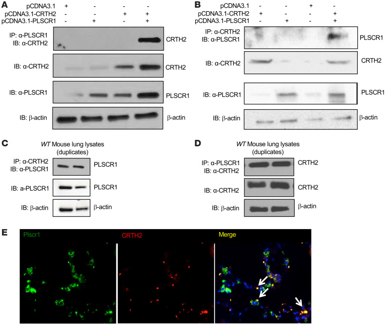
Figure 5. Plscr1 interacts with CRTH2 in vitro and in vivo.
(A and B) HEK293 cells were transfected with CRTH2 and PLSCR1 plasmids and the cell lysates were subjected to either co-IP with α-CRTH2 antibody and immunoblot (IB) with α-PLSCR1, or the opposite. Individual IBs with α-CRTH2 and α-PLSCR1 were also included. (C and D) Mouse lung protein lysates were subjected to either co-IP with α-CRTH2 antibody and IB with α-PLSCR1, or the opposite. Individual IBs with α-PLSCR1 and α-CRTH2 were also included. (E) Lungs from WT mice were sectioned, CRTH2 was labeled with red fluorescence (Alexa Fluor 594) and Plscr1 was labeled with green fluorescence (Alexa Fluor 488). Colocalization of CRTH2 and Plscr1 is indicated by arrows. Nuclei are stained with DAPI (blue). Images are representative of 3 mice.

