Figure 5.
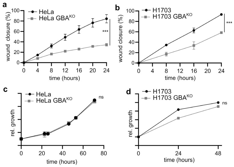
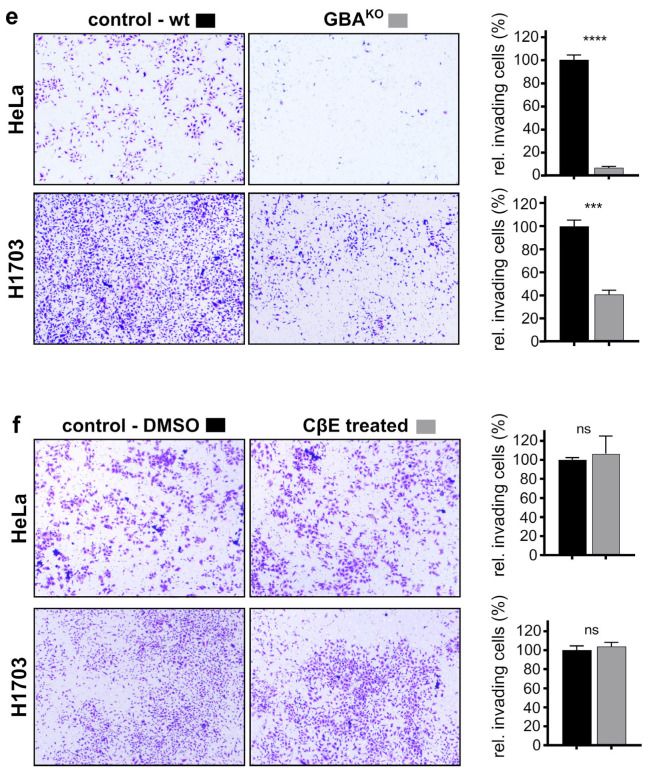
GBA depletion reduced the migratory and invasive capacity of HeLa and H1703 cells, but CβE did not affect their invasive capacity. (a,b) Cell migration in HeLa (a) and H1703 (b) was assayed after a linear segment of confluent cells was removed by scratching. Cells were grown at 37 °C, 5% CO2, and imaged at the indicated times; N = 3 for each line. Wound closure was plotted as the wound closure relative to that measured at t = 0 h. (c,d) The growth of HeLa/HeLa GBAKO (c) and H1703/H1703 GBAKO (d) cells was monitored in 96-well plates using WST-8 assays at the indicated times; N = 5 each line. In (a–d), graphs are scatter plots with the mean ± SEM. ANOVA (Prism): ns, not significant; *** p ≤ 0.005. (e,f) Representative images and corresponding quantification of the indicated cell lines that migrated through a Matrigel matrix and were stained with crystal violet. In (f), cells were treated with either DMSO (0.01% w/v) or 35 µM CβE for 10 days with biweekly medium changes. For all panels, cells were pre-starved for 2 h prior to seeding in the upper chamber of a transwell insert containing Matrigel. Cells were allowed to grow for 48 h at 37 °C and 5% CO2. The lower chamber contained medium supplemented with 10% FBS. Plots are bar graphs with the mean ± SEM; n = 3 for each line. t-Test analysis (Prism): *** p ≤ 0.005; **** p ≤ 0.0001.


