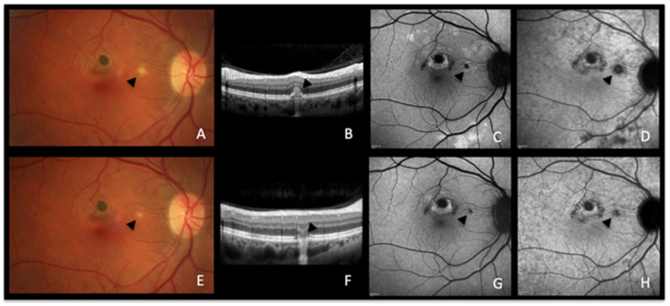
Figure 3.
MFC imaging during active and inactive disease. (A–D) baseline acquisitions. (E–H) acquisitions after recovery. (A,E) colour fundus photographs showing a new fluffy lesion (black arrowhead) (A) evolving to an inactive scar (E). (B,F) optical coherence tomography showing the morphology of the active lesion (B, black arrowhead), recovering in the convalescent phase (F, black arrowhead) with a remaining scar. (C,G) blue-light autofluorescence showing hyperautofluorescent areas in the active stage (C) resolved in the convalescent treated stage (G). (D,H) late indocyanine green angiography showing hypofluorescent areas in the active stage (D), resolved in the convalescent treated stage (H).

