Figure 4.
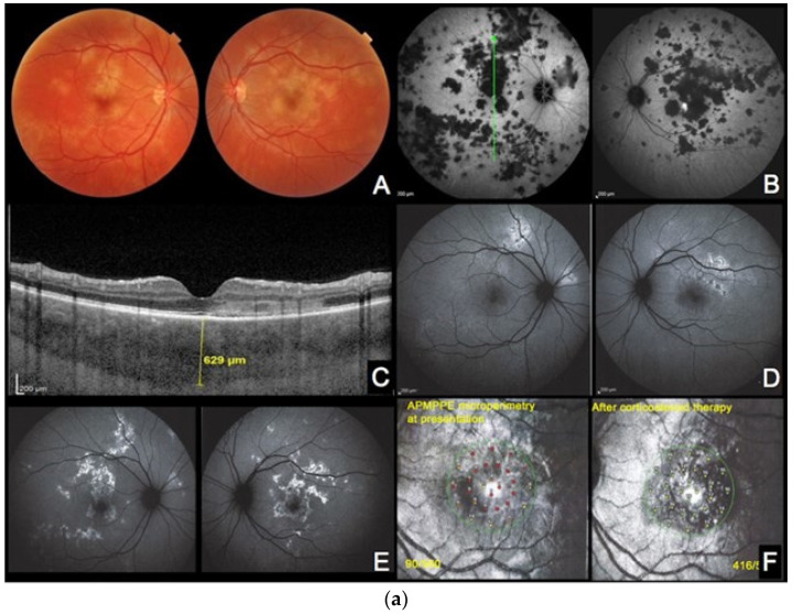
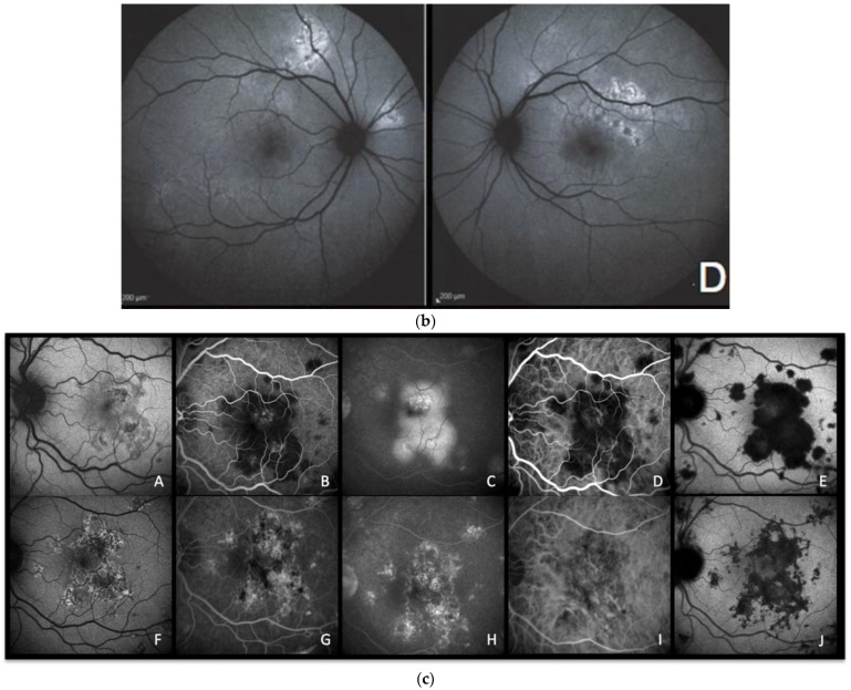
(a) APMPPE during active and inactive disease. (A–D), baseline acquisitions. (E,F) acquisitions after recovery. (A) colour fundus photography showing bilateral yellowish placoid lesions. (B) late-phase indocyanine green angiography showing extensive hypofluorescent areas of choriocapillaris nonperfusion. (C) optical coherence tomography (scan position as shown in (B)) showing oedematous thickening and disorganisation of the outer retina and thickening of whole choroid (629 µm) (D,E) fundus autofluorescence, at the hyperacute stage with minimal hyperautofluorescent areas yet (D), followed by bright hyperautofluorescent areas due to RPE damage at a later stage (E). (F) macular mesopic microperimetry (OS) showing a severe decrease of retinal sensitivity at presentation (90/560), recovering after treatment to 416/560 (bottom far right). (b) APMPPE (enlargement of Figure 4a (A,D)) These BAF frames were obtained at the hyperacute stage of APMPPE when nonperfusion was already widespread (Figure 4a (A,B)) before lesions causing RPE damage were established and apparent on BAF in all areas of nonperfusion. (c) APMPPE during active and inactive disease. (A,E) baseline acquisitions. (F–J) acquisitions after recovery. (A,F) blue-light autofluorescence is quasi-absent at the early stage due to profound ischaemia-induced changes (A) and followed thereafter by bright hyperautofluorescent areas indicating RPE damage (F); (B,G) represent early phase fluorescein angiography (FA) in acute disease showing hypofluorescence due to choriocapillaris nonperfusion (B), while in inactive disease, hyperfluorescent areas indicate window defect due to atrophy (G). (C,H) represent late-phase FA showing in the acute phase pooling due to permeability of retinal vessels induced by severe outer retinal ischaemia (C), while in inactive disease, FA shows the same hyperfluorescent window-defect atrophic areas. (D,E,I,J) represent indocyanine green angiographic frames showing hypofluorescence due to choriocapillaris nonperfusion, early and late, in the acute phase (D,E) and hypofluorescence due to atrophy, early and late, in the inactive phase (I,J).


