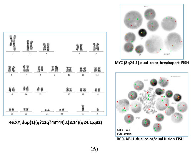
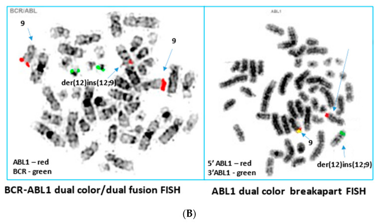
Figure 1.
(A) Representative karyotype from the blood sample obtained at initial diagnosis with Burkitt Leukaemia (left panel), demonstrating a t(8;14) and duplication of 1q. Interphase FISH analysis with a dual colour breakapart probe for the MYC gene (8q24.1) showed MYC gene rearrangement in the majority of cells (upper right panel). After the patient subsequently presented with MPN, BCR-ABL1 FISH performed on an archived Burkitt Leukaemia sample yielded normal results (lower right panel). (B) By G-band analysis of the myeloproliferative neoplasm, the karyotype appeared normal. Sequential BCR-ABL1 metaphase FISH analysis on G-banded cells showed insertion of the ABL1 signal into 12p13, with no involvement of the BCR gene (left panel). Metaphase FISH analysis using an ABL1 breakapart FISH probe (right panel) showed the insertion of 3′ABL1 into 12p13. Results of the ETV6 breakapart as well as the subtelomeric 9q and 12p FISH testing were normal, consistent with an insertion mechanism rather than a translocation mechanism in the generation of the ETV6-ABL1 fusion identified by molecular analysis.


